2D Dental Radiography Interpretation Online Course

Advance your radiographic interpretation skills with Dr. Sholapurkar's in-depth course, tackling dental pathologies and refining diagnostic acumen through interactive case discussions.

8 Chapters

5 hrs

5 CE/CPD
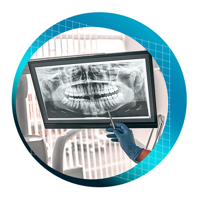
Systematic approach to interpretation of extraoral and intraoral radiographs

Dive deeper into dental radiographic interpretation with Dr. Amar Sholapurkar's 2D Radiography Interpretation course. Building upon the foundational skills, this engaging session delves into dental cases, differential diagnoses, and considerations.

Through the recordings of the interactive discussions and a wealth of clinical images, Dr. Sholapurkar helps participants in refining their diagnostic acumen for pathologies commonly encountered in dental practice.

Suitable for general dentists, this course empowers clinicians to elevate their radiographic interpretation skills and enhance patient care under the instruction of an experienced oral radiology educator.

Course Outline


What will I learn by taking this course?

  • Recognize normal anatomy, differentiating normal from abnormal, and applying skills to clinical scenarios.
  • Focus on interpreting panoramic radiographs, identifying landmarks, air spaces, and employing systematic approaches.
  • Analyze lesions based on location, size, shape, borders, internal structure, and effects on surrounding structures.
  • Learn developmental anomalies like tooth size abnormalities and radiographic patterns of pathologies like cementoblastoma and odontomas.
  • Identify various soft tissue calcifications visible on dental radiographs and give appropriate reference if required.
  • Identify maxillary sinus disorders and cysts/tumors.
  • Recognize and appropriately some uncommon conditions, anatomical variations, and technical errors.
  • Interactive case studies to apply skills in common radiographic interpretation relevant to a general dentists.

Sample the Online Course Content Below!

Get unlimited access to 5 hours of 2D Radiography training, complete with many tips and tricks from an experienced Dental Radiology educator. 

With lifetime access, you can revisit the course whenever you need at your own pace.

All course lessons are available with English subtitles.

Here’s what’s included:
8 Chapters - 17 Video Lessons

CPD/CE Hours: 5

A Foundation for Accurate 2D Dental Radiograph Diagnosis

01 - 01 - Introduction and Overview of Course
  • Recognize normal anatomic landmarks on radiographs, providing a solid foundation for identifying abnormalities.
  • Systematically analyze clinical pictures and radiographs, simulating real-world patient scenarios to enhance diagnostic abilities.
  • Differentiate between normal entities and pathologies, ensuring accurate diagnoses and appropriate treatment planning.
  • Confidently arrive at diagnoses independently, empowering you to make informed decisions in your dental practice.
  • Engaging in interactive discussions and workshops fosters a deeper understanding of radiographic interpretation.
  • Apply the knowledge gained from analyzing over 300 clinical pictures and radiographs to real-world patient cases.
01 - 02 - Normal Dental Radiographic Landmarks and Variants
  • Identify and describe various normal anatomic structures on radiographs, such as the incisive foramen, nasopalatine canal, and pterygomaxillary fissure.
  • Recognize normal variants like the nasolacrimal canal, lateral fossa, and glossopharyngeal air space, preventing misdiagnosis as pathologies.
  • Understand the importance of the pterygomaxillary fissure in diagnosing Le Fort fractures and maxillary sinus pathologies.
  • Differentiate between normal periodontal ligament space variations and pathological widening, ensuring accurate assessment of periodontal health.
  • Integrate radiographic findings with clinical examination and patient history to arrive at accurate diagnoses, emphasizing the role of radiographs as diagnostic aids.
01 - 03 - Differentiating Cervical Burnout from Dental Caries on 2D Radiographs
  • Understand the radiographic appearance and etiology of cervical burnout, recognizing it as a normal radiographic finding rather than a pathology.
  • Identify the key radiographic features that distinguish cervical burnout from dental caries, such as location, shape, and the presence or absence of enamel knife-edge destruction.
  • Integrate radiographic findings with clinical examination techniques, such as probing, to confirm the presence or absence of dental caries.
  • Appreciate the importance of taking multiple radiographs from different angles to avoid misdiagnosis and ensure accurate interpretation.

Navigating Normal Anatomy and Detecting Pathologies on Dental Panoramic Radiographs

02 - 01 - A Systematic Approach to Dental Panoramic Radiograph Interpretation 
  • Understand the importance of knowing normal radiographic anatomy, variants, and common pathologies for accurate interpretation.
  • Differentiate between normal anatomic structures and pathologies by assessing their characteristics, such as bilaterality, symmetry, and location.
  • Identify and describe key normal anatomic landmarks on panoramic radiographs.
  • Recognize and explain the concept of double real images in panoramic radiographs, which occur due to the projection of midline structures.
  • Identify and differentiate various air spaces visible on panoramic radiographs, such as the glossopharyngeal, palatoglossal, and nasopharyngeal air spaces.
  • Apply patient positioning techniques to minimize the appearance of undesirable air spaces, such as the palatoglossal air space, which can obscure important anatomic structures.
02 - 02 - A Case-Based Guide to Dental Panoramic Radiographs

Understand the importance of a comprehensive patient history and clinical examination in guiding radiographic interpretation.

  • Apply a systematic and sequential method for interpreting panoramic radiographs, ensuring that all areas are thoroughly assessed and no pathologies are missed.
  • Recognize the significance of evaluating both dental and non-dental structures in the panoramic radiograph, as the entire image is the responsibility of the dental professional.
  • Identify multiple pathologies in a single radiograph by assessing the entire image rather than focusing solely on the area of interest.
  • Appreciate the role of histopathological examination as the gold standard for definitive diagnosis, while recognizing the value of radiographic interpretation in providing differential diagnoses.
  • Understand the importance of reviewing images in a relaxed state to minimize the risk of overlooking important findings.

Radiographic Diagnosis of Oral Lesions and Anomalies

03 - 01 - Decoding Dental Radiographic Lesions
  • Determine whether a lesion is odontogenic or non-odontogenic based on its location relative to the inferior alveolar nerve canal or the maxillary antrum.
  • Understand the significance of a lesion's site and location in narrowing down the differential diagnosis.
  • Assess the size of a lesion using measuring tools and describe its dimensions in millimeters or centimeters.
  • Analyze the shape of a lesion and understand how it relates to the likelihood of various pathologies, such as cysts, tumors, or abscesses.
  • Evaluate the borders of a lesion and recognize how different border characteristics, such as corticated, sclerotic, or ill-defined borders, are associated with specific pathologies.
03 - 02 - Internal Structures and Surrounding Effects - Lesion Analysis
  • Describe the internal structure of lesions using terms such as radiolucent, radiopaque, or mixed, and recognize how these characteristics relate to specific pathologies.
  • Identify the presence of septae or calcifications within lesions and understand their diagnostic significance.
  • Recognize the effects of lesions on surrounding structures, such as bone resorption or formation, and interpret these changes in the context of inflammatory diseases.
  • Understand how space-occupying lesions create their own space by displacing adjacent structures, such as teeth, the maxillary sinus, or the inferior alveolar nerve canal.
  • Identify root resorption and relate this finding to the presence of trauma, cysts, or slow-growing benign tumors.
  • Recognize cortical bone expansion as a characteristic feature of benign, slow-growing tumors or cysts.
  • Apply a systematic approach to radiographic interpretation using the "4S and IE" method, which includes assessing site, size, shape, surface (borders), internal structure, and effects on surrounding structures.

Common Radiographic Spectrum of Dental Pathology

04 - 01 - Radiographic Diagnosis on Developmental Anomalies and Clinical Implications 
  • Recognize and differentiate between microdontia, macrodontia, and other tooth size abnormalities on radiographs.
  • Identify supernumerary teeth, such as mesiodens, distomolars, and paramolars, using specific terminology to describe their location and characteristics.
  • Understand the radiographic appearance and clinical implications of fusion, gemination, and taurodontism.
  • Apply a systematic approach to diagnosing developmental anomalies, such as dens invaginatus, through a combination of thorough clinical examination and radiographic interpretation.
  • Recognize the radiographic features of dens evaginatus, talon cusp, and Turner's hypoplasia, and understand their clinical significance.
  • Identify concrescence on radiographs and appreciate its importance in treatment planning, particularly for extractions.
  • Recognize the radiographic appearance of dilacerated roots in both the anteroposterior and buccolingual directions, and understand the implications during extractions.
  • Differentiate between odontogenic and non-odontogenic lesions, such as Stafne's defect, based on their location and radiographic characteristics.
04 - 02 - Radiographic Patterns of Dental Pathology
  • Recognize erosion on radiographs and differentiate it from positioning errors in panoramic radiography.
  • Identify periapical cemental dysplasia and understand its radiographic stages.
  • Differentiate between condensing osteitis and hypercementosis and other periapical radiopacities.
  • Recognize enostosis (idiopathic osteosclerosis) and distinguish it from exostosis or tori through clinical examination and radiographic interpretation.
  • Identify the characteristic radiographic features of benign cementoblastoma, including its association with vital teeth and the presence of a soft tissue capsule.
  • Differentiate between compound odontoma, complex odontoma, and dilated odontoma based on their radiographic appearance, location, and clinical presentation.
  • Recognize the unique radiographic presentation of dilated odontoma, such as the "donut" appearance when present in the mandible.
  • Distinguish between root fragments and bony spicules on radiographs by assessing the presence of the periodontal ligament space and pulp canal.

Dental Radiographic Detection of Soft Tissue Calcifications

05 - 01 - Soft Tissue Calcifications From A Dental Radiographic Perspective
  • Differentiate between heterotopic calcification and heterotopic ossification, and recognize their radiographic appearances.
  • Identify normal anatomical structures that may appear calcified on panoramic radiographs, such as the thyroid cartilage and triticeous cartilage.
  • Recognize the radiographic features of calcified lymph nodes, which often resemble a cauliflower or coral-like appearance.
  • Understand the importance of eliciting a thorough patient history to identify potential causes of lymph node calcification, such as granulomatous disorders or previous infections.
  • Identify tonsilloliths on panoramic radiographs and correlate their location with the glossopharyngeal airspace overlapping the ramus of the mandible.
  • Recognize the clinical features and symptoms associated with tonsilloliths, such as pain, swelling, halitosis, and foreign body sensation.
  • Differentiate tonsilloliths from other soft tissue calcifications, such as parotid calculi, based on their location and associated clinical findings.
  • Understand the strategies for identifying tonsilloliths, including observation, referral for surgical removal, and the importance of follow-up in certain patient populations.
05 - 02 - Diagnosis of Calcifications seen in Dental Panoramic Radiographs
  • Recognize the radiographic appearance of calcified atherosclerotic plaques and understand their location in relation to anatomical landmarks such as the greater cornu of the hyoid bone and cervical vertebrae.
  • Appreciate the importance of referring patients with suspected calcified atherosclerotic plaques to a physician for further evaluation and management, as they may be at risk for stroke.
  • Differentiate calcified atherosclerotic plaques from other calcifications, such as the triticeous cartilage, based on their location, shape, and homogeneity.
  • Identify sialoliths on radiographs and correlate their presence with clinical symptoms such as pain and swelling in the floor of the mouth or parotid region.
  • Understand the importance of reducing exposure when imaging suspected sialoliths due to their potential low mineral content.
  • Recognize the radiographic appearance of phleboliths and their association with hemangiomas, as well as the clinical implications of these lesions during surgical procedures.
  • Differentiate between antroliths and rhinoliths based on their location within the maxillary sinus or nasal cavity, respectively, and understand their typical clinical presentations.
  • Develop a systematic approach to identifying various soft tissue calcifications encountered in dental practice, including when to refer patients to GP or specialists for further evaluation and treatment.
05 - 03 - Presentations of Stylohyoid Ligament Ossification
  • Identify the radiographic appearance of stylohyoid ligament ossification on panoramic radiographs.
  • Differentiate between the three main aspects of stylohyoid ligament ossification: Styloid Chain Ossification, Eagle Syndrome, and Stylohyoid (Carotid Artery) Syndrome.
  • Understand the clinical significance of each aspect and the associated signs and symptoms that patients may present with.
  • Recognize the importance of thorough patient history-taking and questioning in arriving at the correct diagnosis.
  • Understand the pathophysiology behind Eagle Syndrome, which involves impingement of the ossified styloid process on the glossopharyngeal nerve, leading to pain and discomfort.
  • Recognize the symptoms associated with Stylohyoid (Carotid Artery) Syndrome, such as otalgia, tinnitus, temporal headache, and vertigo, and understand the mechanism of sympathetic nerve tissue irritation in the carotid artery wall.
  • Develop a systematic approach to analyze patients with stylohyoid ligament ossification, including when to refer patients to ENT specialists for further evaluation and treatment.
  • Brief understanding of surgical identification of Eagle Syndrome and Stylohyoid (Carotid Artery) Syndrome, which involves amputation of the ossified styloid process.

Radiographic Diagnosis of Maxillary Sinus Pathology and Odontogenic Tumors

06 - 01 - A Radiographic and Clinical Approach to Diagnosing Maxillary Sinus Disorders
  • Recognize the radiographic appearance of maxillary sinus mucositis and understand its clinical implications and management.
  • Identify periostitis of the maxillary sinus floor on radiographs and correlate its presence with periapical inflammation of maxillary posterior teeth.
  • Understand the concept of maxillary sinus pneumatization and its impact on treatment planning, particularly for implant placement.
  • Recognize the radiographic features of retention pseudocysts in the maxillary sinus and differentiate them from other sinus pathologies based on clinical presentation and patient symptoms.
  • Identify the radiographic appearance of mucoceles in the maxillary sinus and understand their etiology, clinical presentation, and potential complications, such as the destruction of the pterygomaxillary fissure.
  • Develop a systematic approach to diagnosing maxillary sinusitis based on clinical findings, such as pain on percussion of maxillary posterior teeth, vitality testing, and the absence of other dental pathologies.
  • Understand the importance of additional diagnostic tests, such as palpation and head tilt, in confirming the diagnosis of maxillary sinusitis.
  • Recognize when to refer patients with maxillary sinus disorders to ENT specialists or general physicians for further evaluation and management.
06 - 02 - Radiographic Interpretation of Cysts and Tumors
  • Recognize the radiographic appearance of odontogenic cysts and tumors, particularly ameloblastoma, and differentiate them from other lesions.
  • Understand the importance of histopathology as the gold standard for definitive diagnosis of cysts and tumors.
  • Identify the characteristic radiographic features of ameloblastoma, such as multilocular appearance and soap bubble-like patterns.
  • Distinguish between odontogenic and non-odontogenic lesions based on their location relative to the inferior alveolar nerve canal.
  • Recognize the importance of referring patients with suspected odontogenic tumors to oral and maxillofacial surgeons for a comprehensive evaluation and appropriate treatment planning.
  • Apply the knowledge gained from previous lessons, such as identifying stylohyoid chain ossification and determining the need for referral based on patient symptoms.

Strategies for Identifying Uncommon Findings and Radiographic Rarities

07 - 01 - Navigating Uncommon Findings and Variations on Radiographs
  • Recognize and differentiate between internal and external root resorption on radiographs, understanding their etiologies and clinical implications.
  • Distinguish between pathological conditions and normal follicular space around impacted canines, ensuring accurate diagnosis and treatment planning.
  • Identify radiographic signs of metastatic carcinoma in the condyles and posterior mandible, particularly in patients with a history of breast cancer in females or prostate cancer in males.
  • Understand the importance of thorough history-taking and targeted questioning in diagnosing metastatic lesions, even in asymptomatic patients.
  • Appreciate the significance of prompt referral to a general practitioner or specialist when metastatic lesions are suspected based on radiographic findings and patient history.
  • Recognize and understand the clinical implications of anatomical variations such as bifid and trifid mandibular canals, which may complicate dental procedures like extractions and implant placement.
  • Identify bifid mandibular condyles on radiographs and understand their potential to mimic fractures or tumors, emphasizing the importance of knowledge and awareness of this entity.
  • Analyze and identify technical errors in radiographs, such as double exposure due to patient movement, ensuring optimal image quality for accurate diagnosis.
07 - 02 - Radiographic Oddities and Terminologies - Unusual Cases and Proper Communication
  • Identify and interpret unusual radiographic findings, such as retained foreign bodies (e.g., gunshot pellets) and surgical artifacts (e.g., gold wire from facelift procedures), and understand the importance of thorough patient history-taking in such cases.
  • Recognize cultural practices that may result in unusual radiographic findings, such as the use of susuk or charm needles in some Southeast Asian countries, and appreciate the importance of cultural awareness in providing patient care.
  • Analyze and interpret radiographic anomalies, such as buccolingually impacted teeth, kissing molars, and multiple impacted teeth, and understand their potential associations with syndromes like Apert syndrome, Crouzon syndrome, or cleidocranial dysplasia.
  • Identify artifacts and errors in radiographs caused by patient movement or the presence of foreign objects, such as mobile phones, emphasizing the importance of proper patient preparation and instruction.
  • Understand and apply correct radiographic terminology in professional communication, such as using "radiograph" instead of "X-ray" and describing findings as radiolucencies or radiopacities rather than directly inferring pathology.
  • Recognize and use appropriate terminology for different radiographic views and techniques, such as distinguishing between a periapical radiograph (IOPA) and a posterior-anterior (PA) view, and using non-proprietary names for panoramic radiographs (e.g., PAN, OPT).

Dental Radiograph Interpretation Workshop

08 - 01 - Honing Your Dental Radiographic Interpretation Skills Through Interactive Case Studies
  • Identify and diagnose a wide range of pathologies, anomalies, and normal anatomical structures on radiographs, such as residual cysts, bifid mandibular canals, tonsilloliths, and soft tissue calcifications.
  • Differentiate between various periapical radiolucencies, such as periapical cemental dysplasia, condensing osteitis, and enostosis, based on radiographic appearance and clinical findings.
  • Recognize and analyze less common conditions, such as lateral periodontal cysts and cementoblastoma.
  • Apply a systematic approach to radiographic interpretation, carefully analyzing all aspects of the radiograph and considering patient history and clinical findings to arrive at accurate diagnoses.
  • Demonstrate the ability to identify and interpret subtle radiographic findings, such as partial stylohyoid chain ossification, emphasizing the importance of thorough and meticulous radiographic examination.
  • Understand the significance of radiographic interpretation in the context of comprehensive patient care, recognizing that radiographs serve as a diagnostic aid and should be used in conjunction with clinical information and other tests to formulate the final diagnosis.

Unveiling the Secrets of Dental Radiography


Dive deeper into dental radiographic interpretation with Dr. Amar Sholapurkar's 2D Radiography Interpretation course.

Learn Different Features

Learn different radiographic features of maxillary sinus disorders, soft tissue calcifications, and more

Systematic Case Navigation

Navigate common cases through systematic, step-by-step radiographic analysis

Differentiate Similar Entities

Confidently differentiate between similar-appearing entities using key diagnostic criteria

Understand Clinical Implications

Understand the clinical implications and considerations for various radiographic findings

Interactive Knowledge Application

Engage in interactive case discussions to solidify knowledge application and critical thinking skills

Learn from an Experienced Educator

Learn from a seasoned oral radiology educator with extensive teaching experience

2D Dental Radiography interpretation

Online Course

$495 USD

  • 8 Chapters
  • 5 hrs of content
  • 4 quizzes to test your understanding
  • 5 CPD Credits
  • Lifetime access!

Buckets of Material

"WOW!!! A lot of information, but as I have come to learn, you have to know your materials. Thanks for a concise presentation."

Summarised it amazingly.

"Tried to make my own summary for hours on end. Found this course which summarised it amazingly in 20mins. Appreciate it so much."

Murshidah Kafrawi

Dental Student

"

You are Fully Protected by Our 100% Satisfaction-Guarantee.

I am confident that you will love this course, if you purchase the course and are not satisfied with the quality or content of the training, then let me know within 30 days of your purchase and I will refund you in full.

No questions asked.

Dr Ahmad Al-Hassiny

Founding Director of the Institute of Digital Dentistry


Meet Your Educator


Dr Amar Sholapurkar

BDS, MDS (Oral Medicine and Radiology)

Dr. Amar Sholapurkar is a highly accomplished academician and clinician with extensive global experience in dental radiology and education. Currently serving as discipline lead of Oral Radiology and Senior Lecturer (Clinical Dentistry and Oral Radiology) at the College of Medicine and Dentistry, James Cook University (JCU), Cairns, Australia, he has garnered numerous accolades for his outstanding contributions to teaching and student learning.

In 2021, he received the JCU Dean's Learning & Teaching Award, and in 2022, he was honored with the James Cook University Citation for Outstanding Contribution to Student Learning Award. He is currently doing a PhD at James Cook University, Australia.

With over 17 years of teaching and research experience in dental radiology, Dr. Sholapurkar has published extensively. He has authored over 50 articles in peer-reviewed dental journals, contributed 11 book chapters, and edited "Publish and Flourish — Practical Guide for Effective Scientific Writing." He has delivered guest lectures and keynote addresses at dental conventions, specialty conferences, and continuing professional development activities in Australia and abroad.

Additionally, he is involved in running Cone-beam Computed Tomography (CBCT) licensing courses and OPG/Lat Ceph licensing courses across Australia, contributing to the professional development of dental practitioners. He also regularly runs CBCT and 2D Radiology interpretation courses.

Dr. Amar Sholapurkar's courses has been verified by the New Zealand Dental Association and can be seen on their website.

Outcome Summary

If you are a New Zealand dental practitioner, this course has been verified by the New Zealand Dental Association and can be seen on their website.

Dr. Sholapurkar delivers this course in an interactive format so this is a recording of one of his live courses so you will feel like you were there with the group.

This highly interactive session incorporates over 300 radiographs and clinical pictures, with each case/pathology having a take-home message.

After the first seven chapters, the eighth chapter is a recording of interactive case studies between Dr. Sholapurkar and delegates using a completely different set of radiographs pertaining to the previously discussed pathologies.

You will be granted 5 CPD hours after passing the four multiple choice quiz tests included with this course with a passing grade of 80%.

>