Artificial Intelligence has been one of the biggest hype topics in dentistry in the past couple of years.
You can see it being discussed constantly at many of the biggest conferences and congresses worldwide.
What is all the fuss about? - Well, it may change how we practice medicine and dentistry completely.
Platforms like ChatGPT have pushed the concepts of AI into the mainstream, and we have seen many industries contemplate how AI can increase efficiency, accuracy, and productivity - dentistry is no different.
When we attended IDS 2023 in Cologne, Germany, we were absolutely blown away by how many useful AI tools have been developed for the dental industry. I think back to IDS 2019, a time when there was literally no one talking about AI in dentistry. Fast-forward a short 4 years, and we are in for a big change soon, it seems.
One really interesting piece of technology is Diagnocat. We have had the pleasure of testing extensively in our group of practices in Wellington, New Zealand for the past 12 months. One of my associates, Dr. Byron Park, has been prolifically using Diagnocat. Together we put all our insights about this interesting tech below, so let's dive into an in-depth breakdown of the pros, cons and benefits of Diagnocat.
Enjoy!
Download the Full Review PDF
Download the PDF to get a full copy
of this article to read later.
Get a high resolution printable copy of this review.
Introduction
At our clinic in Wellington - the home of the Institute of Digital Dentistry - we’re always proud to test exciting and innovative technology. So, we could hardly say no when Diagnocat reached out for us to be the first clinic in the Australiasia region to try out Diagnocat!
This is one of the first AI software in the world to completely focus on dentistry and, in particular - CBCT images. We have put through hundreds of cases through this software and now feel ready to share our objective review with you.
Disclaimer - no conflict of interest. This is an objective review of Diagnocat. The team at iDD remains unwaveringly committed to providing you with impartial and trustworthy information. Diagnocat had no part in writing this review or restricting any conclusions iDD makes in our thorough analysis and clinical use of this software.
What is Diagnocat?
In short, Diagnocat is a powerful AI service that analyzes dental images and provides you with a report.
That is fundamentally what it does.
These reports are generated from X-rays and include 3D CBCTs, and 2D images like panoramic X-rays, bitewings, and periapical radiographs.
Much like other AI services in dentistry, it is completely web-based and utilizes cloud computing. In simple terms, you upload any x-ray to the Diagnocat website, it takes a minute or two to read it, and then generates a report.
You can either manually upload X-rays to the platform or have it automatically sync with your practice management system, meaning that anytime you take an X-ray it automatically uploads it to the Diagnocat platform.
Some people are worried about the ramifications of AI, especially in healthcare. It is important to understand that AI is not replacing diagnosis by clinicians. This is totally the wrong thinking.
The company positions Diagnocat as a trusted virtual assistant. It simply generates a completely objective report based on the radiographs, and it is your choice what to do with that information.
In this light, AI can help improve case acceptance and clinical efficiency, especially when presented to patients, as it is completely objective and never ‘upselling.’ AI also helps standardize care between clinicians.
We were lucky enough to get an introduction about the company and multiple training sessions on Diagnocat by the CEO and creator, Alex Sanders.
Alex created Diagnocat because he owned a network of dental clinics with more than 100 dentists - all with varying experience and abilities. This was a very big company, and it became evident that the ability to diagnose radiographs varied considerably among dentists. This was a problem when it came to consistency of care for patients.
With this in mind, Alex created the Diagnocat in 2017 and developed it into what it is today. The software was trained on a dataset of over 35,000 radiographs and expanded to include 3D CBCT radiographs and other services.
What services does Diagnocat offer?
Diagnocat offers radiological reports for intraoral X-rays (PBWs and PAs), OPG, and CBCT. Your trusty advisor, in a way.
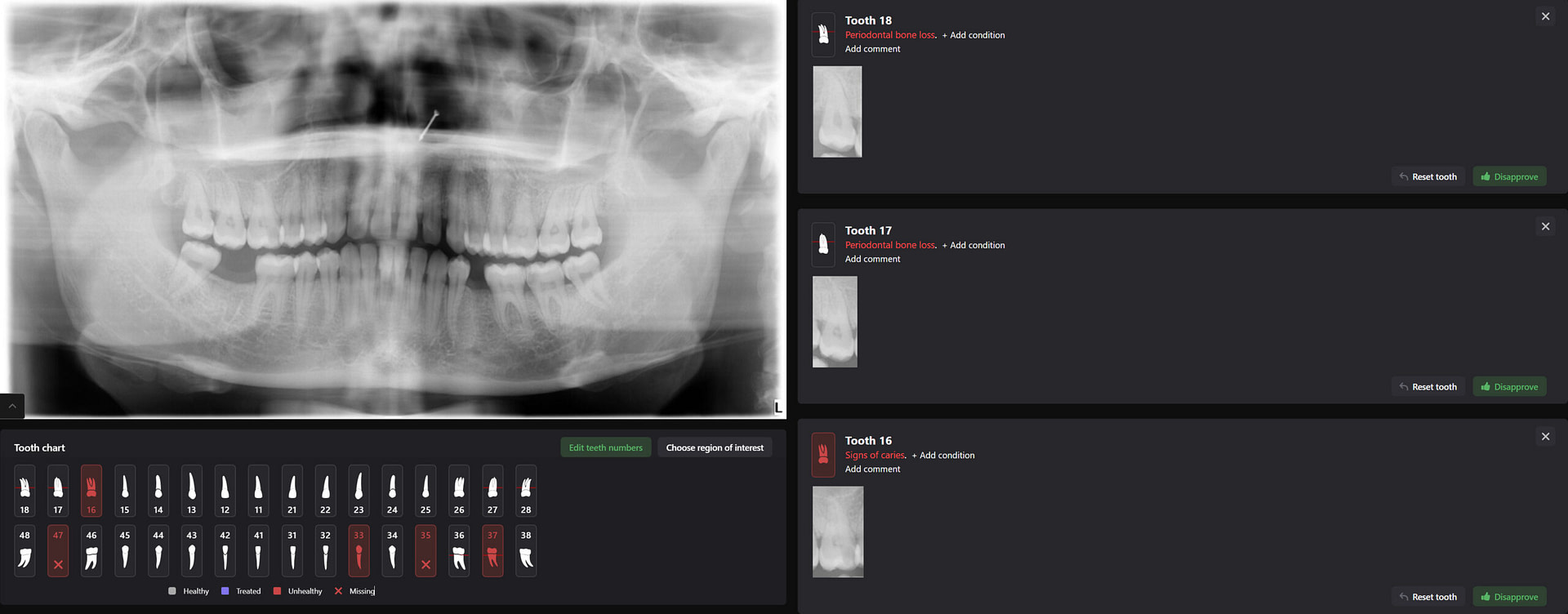
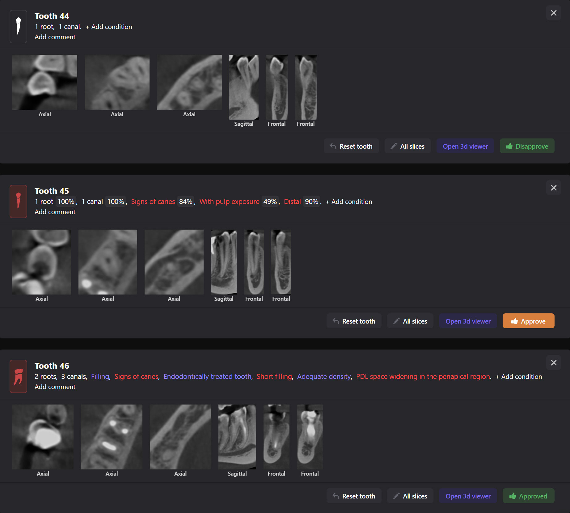
Every tooth in the radiograph is identified by its ISO (FDI) or Universal (American) number automatically. It can even pick up missing teeth. This is all labeled and identified in its intelligent reporting system.
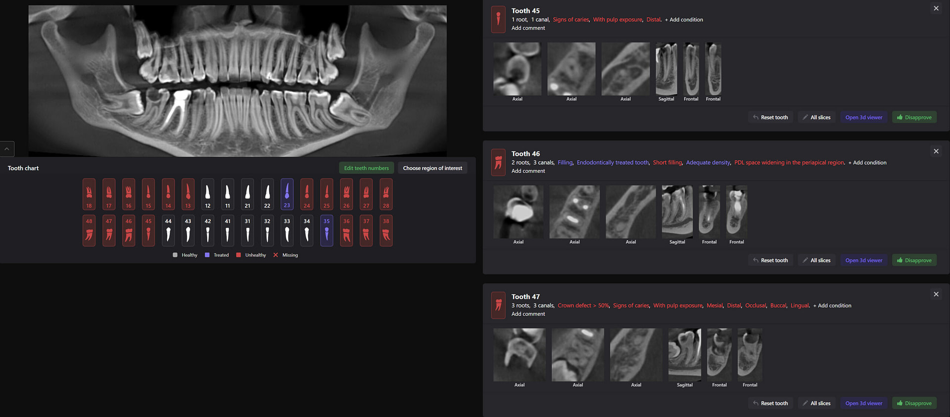
A report is generated and outlines the health of every single tooth. For each tooth, Diagnocat can identify 25 different possible findings for 2D images and more than 65 for 3D radiographs. Each tooth report can be approved, and the dentist’s signature can be added to the overall report, which adds accountability to the report's contents.
What is AI Diagnosis?
AI diagnosis is the use of artificial intelligence techniques and algorithms to aid in the process of diagnosing medical conditions or diseases. It involves machine learning, deep learning, and other AI technologies to analyze patient data, medical images, laboratory results, and other relevant information to support medical professionals in making accurate and fast diagnoses.
At the time of this article, several available systems are capable of AI diagnosis of dental radiographic images, including:
- Pearl - Second Opinion®
- Overjet
- VELMENI - Second Dentist™
- ORCA Dental AI
- Denti.AI
- Diagnocat
- Videa Health
- adravision
- AI:Dental
- dentalXrai
- Allisone
One of the things that makes Diagnocat stand out is that most other AI software can only do 2D images. This is true for the vast majority of the list above. In comparison, Diagnocat can do both 2D and CBCT diagnosis.
How has using Diagnocat benefited our clinic?
Wow factor
One of the hardest things to measure ROI for when using digital tools is the wow factor and the dividends it pays.
Patients were genuinely impressed (in many cases blown away) when I explained that AI reads their X-rays and generates a report. The speed at which the report is created and how thorough it is with more than 65 conditions that can be diagnosed is extremely impressive to patients. As mentioned above, we are in a time now that AI is in our daily language. There are news reports on ChatGPT every month. The average person understands how powerful AI is and how it is changing the world around us.
Speed
There is no point in using AI diagnostics if it takes longer than you to read an x-ray.
Generating a report using Diagnocat for a full mouth series of up to 24 2D radiographs (PAs, PBWs) takes less than a minute. Yes, it is impressively fast.
Generating a full report of a full-mouth CBCT 3D radiograph takes four to six minutes. We are talking about going over every slice with a fine-tooth comb. This has huge benefits, as a maxillofacial radiologist may take up to 40 minutes to fully report on a 3D CBCT radiograph.
These reports can be generated in the background while the clinician performs the intraoral examination. The dentist can then show the report in real-time in the post-examination discussion with the patient.
Once again, the report supplements your own diagnoses and clinical findings.
Reducing errors/missed findings
AI doesn’t have an off day where it misses a morning coffee. Nor an exhausting day after a tough wisdom tooth extraction. Or even a tiring afternoon after your fifth new patient examination.
It will consistently pick up findings. That does not mean it is 100% accurate. However, the company claims that based on their data, it has screening accuracy up to 90%. Basically, it rarely misses, but you still have to check the report carefully.
Diagnocat can also detect many more ‘shades of grey’ on an x-ray than the human eye can see. It can compensate for borderline unreadable radiographs that were over/underexposed. And it frankly knows more diagnoses on a CBCT than some (maybe many) dentists. Remember - 65 different things it's looking for. For fun, try to write down 65 possible diagnoses that can come up on a CBCT.
Treatment plan acceptance
Artificial intelligence is seen as a neutral third party that has nothing to benefit from the findings. In our experience, patients readily accept the diagnosis if you can explain how the AI picked up the finding.
If you can then guide the patient to understand the detrimental effects of not treating the condition, they generally will want to do something about it. I have noticed an increase in the uptake of dental treatment that has been planned using Diagnocat.
We all know as dentists that our patients sometimes make comments like ‘I bought your next holiday’ or car etc. Some patients do not trust us and may see this as a conflict of interest. They can feel like dentists are up-selling. With AI this is just simply not the case. People trust it.
CBCT segmentation
This is one feature that actually made Diagnocat very popular early on. It was the only software that made CBCT segmentation easy. Nowadays, there are several different companies and software that carry out segmentation. Diagnocat was the first and is still arguably the best at it.
So what is Segmentation? It basically means taking the CBCT and delineating all the different 3D structures, bones, individual teeth, etc.
These can then be individually exported via the software by generating STL files from CBCT dicom data. This can be used in other dental software. Jaw STLs can be used in Modjaw (jaw motion capture) for TMJ analysis, for example. As well as in exocad to help visualize the jaws for surgical planning or guide creation.
Being able to take a DICOM file and turn it into STLs is useful for a host of different CAD/CAM indications.
Download the Full Review PDF

Download the PDF to get a full copy
of this article to read later.
Get a high resolution printable copy of this review.
Real clinical examples of using Diagnocat
Upon reflecting on our use of Diagnocat in our clinical practice, I feel like the best way to show you the benefits is to describe some clinical situations that come to mind.
- I have inherited patients from a retired colleague whose methods, though effective in their time, may not reflect the most current standards of care. As a result, many of his patients had extensive dental disease despite decades of yearly examinations/cleans with him. This was a tricky situation to navigate, as I would never throw a colleague under the bus. However, the patients needed to be informed of their conditions. From the patient’s perspective, I am a new dentist with whom they have yet to establish trust and a relationship. Diagnocat helped me as a neutral observer to diagnose the problems in their mouth and allowed me to discuss them without seeming to have a motive.
- There have been instances in the past where I have taken x-rays in a check-up appointment and discussed a treatment plan which the patient has accepted. After the patient leaves, I typically write my clinical notes and recheck their X-rays. Occasionally, I find something else I did not pick up when the patient was there. These new findings may affect the treatment plan. The patient needs to be informed of these and a new agreement made. Diagnocat has helped to reduce this problem as generally findings are picked up very well. It has especially amazed me how well it can detect very subtle findings, such as early interproximal and occlusal radiolucencies.
- I had a severely dentally anxious patient with a previous traumatic dental history. Supposedly a dental assistant had to hold her down during a painful root canal procedure. This had left a lasting traumatic mental scar, and the patient would not even sit in the dental chair for an examination. However, she consented to taking a 3D CBCT radiograph and AI diagnosis. The radiograph and report were so clear I was confident in making her entire treatment plan from this. Of course, this would not be considered ideal treatment planning without an intraoral examination. However, real-world situations like this demand our empathy and flexibility. Her treatment was then completed under intravenous sedation.
- CBCT segmentation
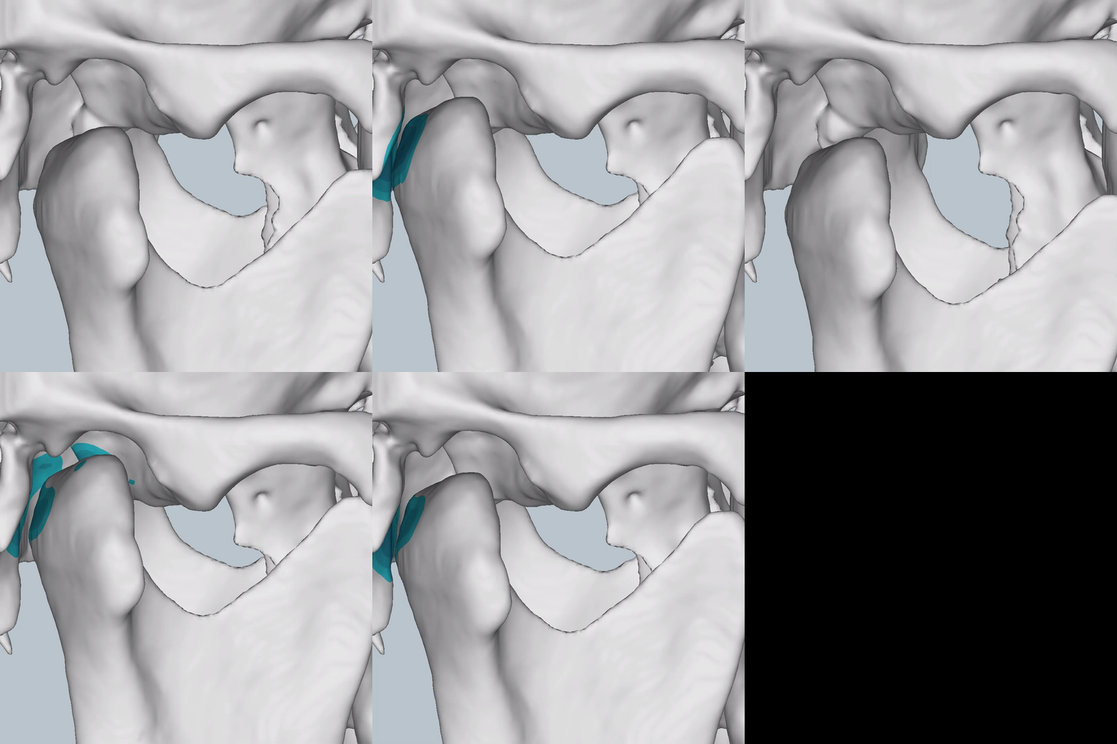
STLs of upper and lower jaws imported into Modjaw for TMJ analysis in jaw motion. this is made possible by CBCT segmentation using Diagnocat.
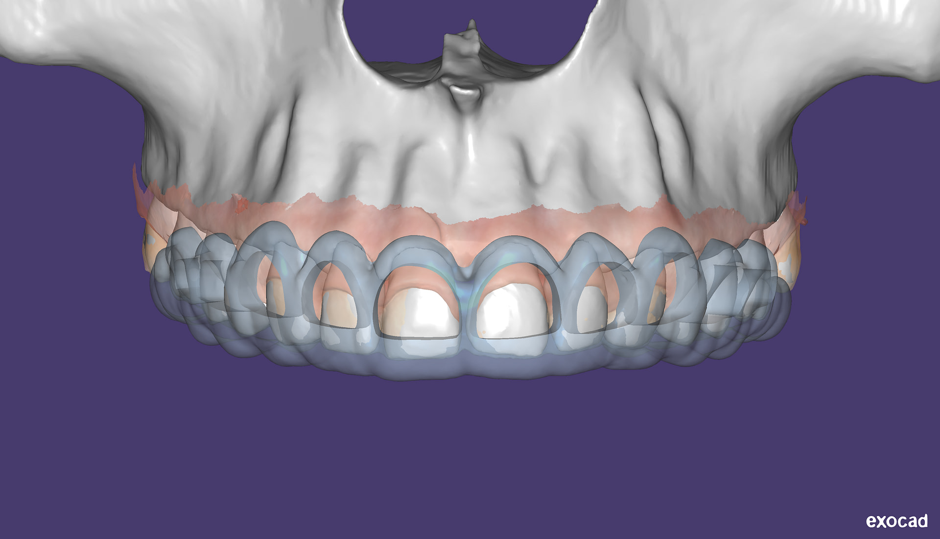
The surgical crown lengthening guide was made with the aid of maxilla STL to visualize crestal bone level. Again thanks to CBCT segmentation by Diagnocat.
Intraoral radiological report
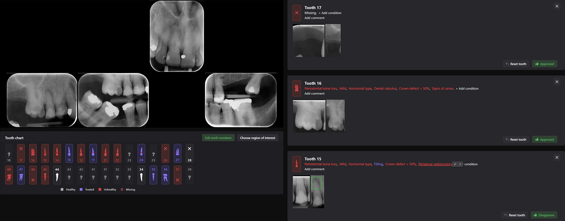
Panoramic radiological report
CBCT radiological report
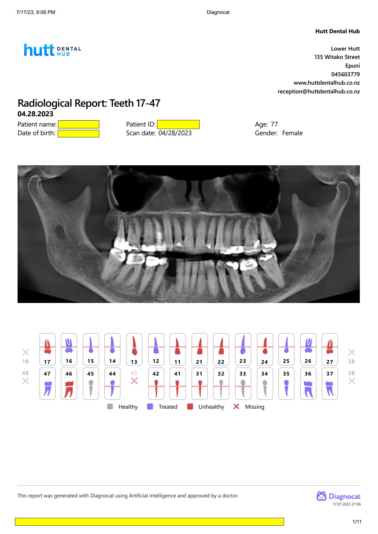
Printed Radiological Report
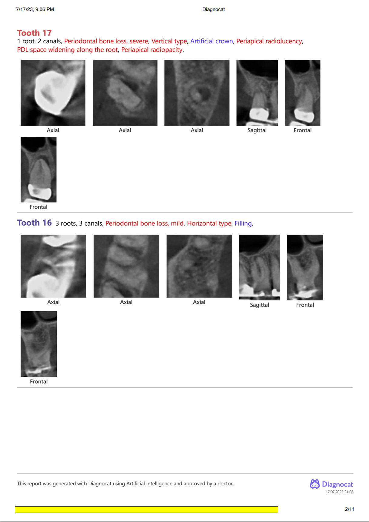
Diagnocat can pick up non-dental pathology as well, such as thickening of the sinus lining and bone cysts.

Orthodontic Report
Diagnocat is also capable of generating orthodontic reports.
This requires a CBCT radiograph with a minimum field of view of 13 x 15 x 15cm. Our Kavo OP3D machine achieves this by taking a double-field exposure and stacking two exposures on top of each other.
Diagnocat generates OPG and frontal and lateral cephalometric reconstructions from the CBCT data.
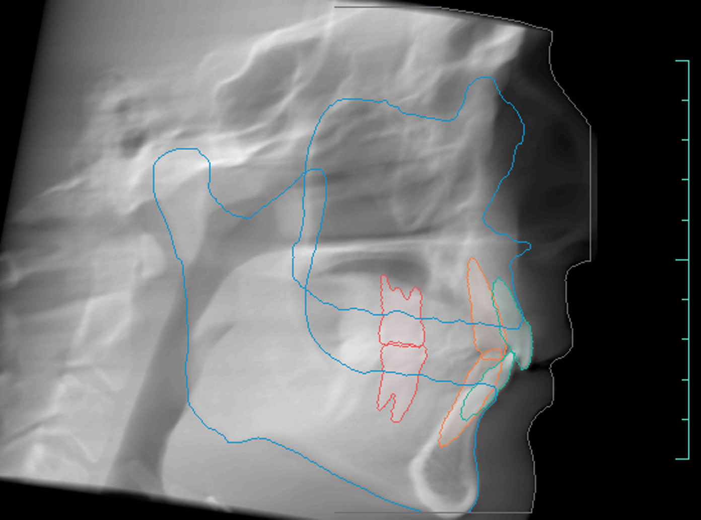
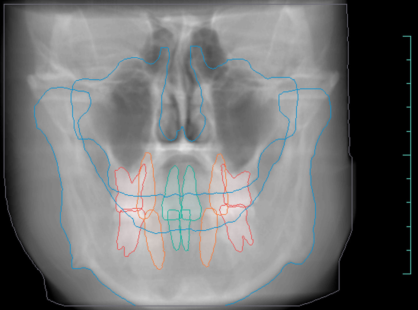
Tracings of the maxilla, mandible, central incisors, canines and molars. All automatically.
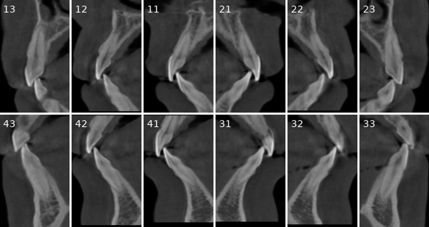
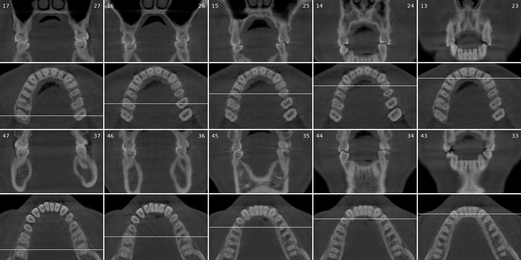
Cross-sectional and coronal views of teeth show torque and buccolingual relationships such as crossbite.
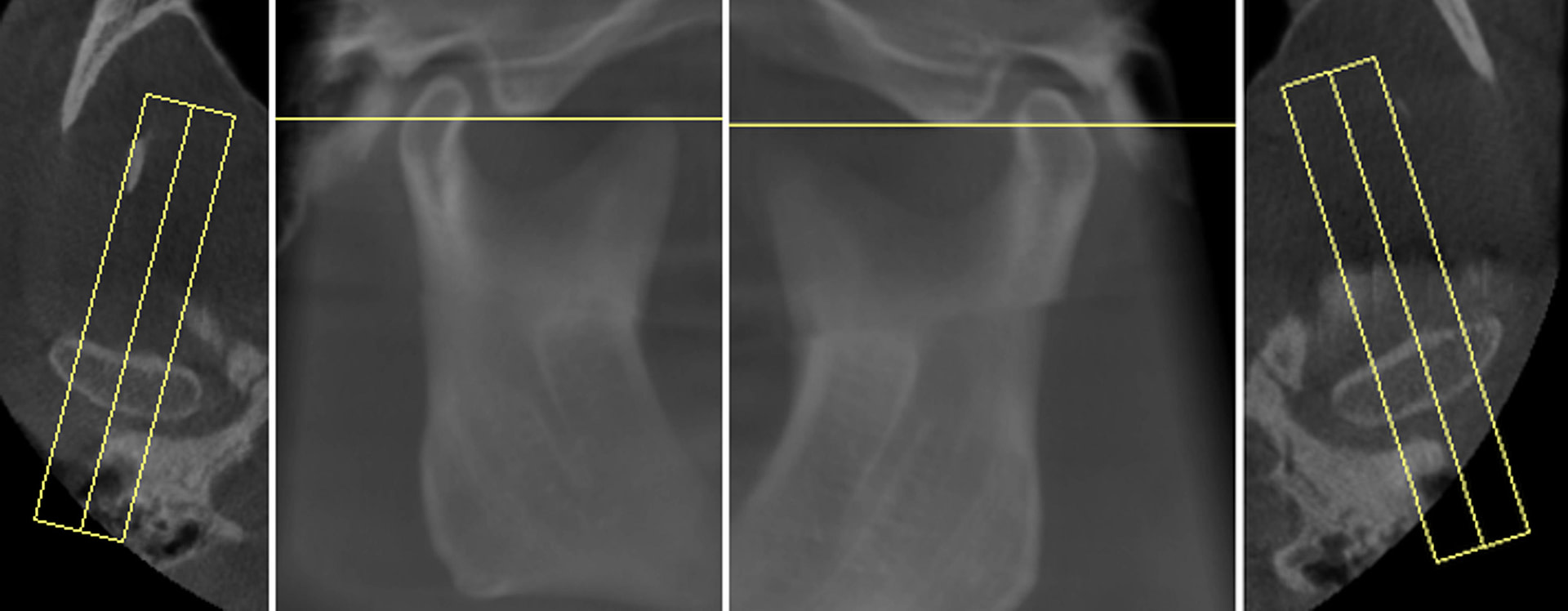
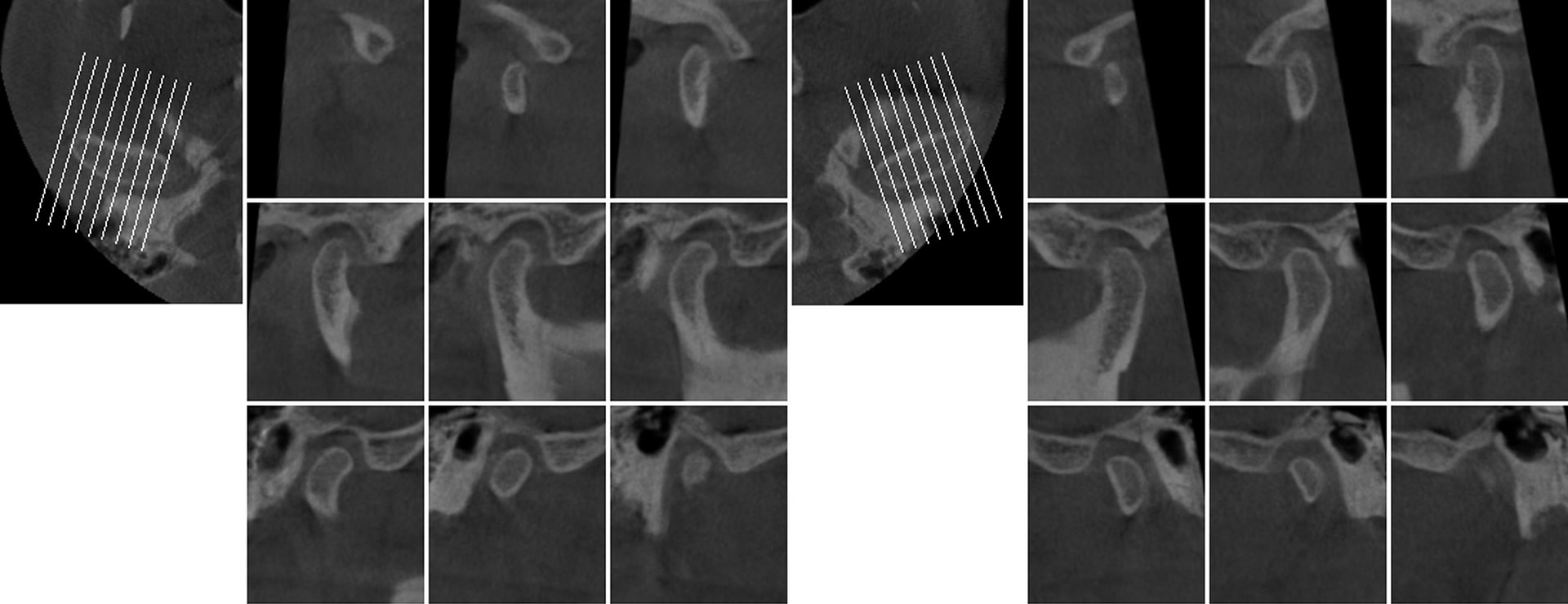
TMJ coronal/sagittal slices and summations visualize abnormal positioning/shape of the mandibular condyle.
Endodontic Report
Tooth number is selected to analyze the root canal morphology within the CBCT radiograph.
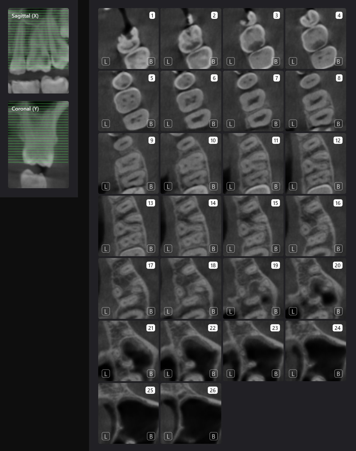
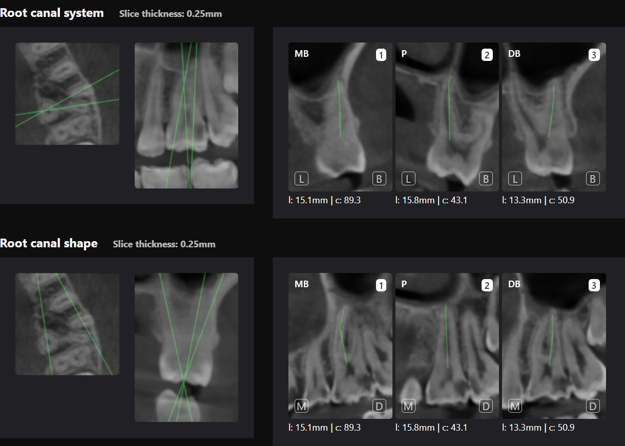
Sagittal and coronal slices and highlighting the angle/curvature/length of the canals.
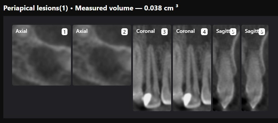
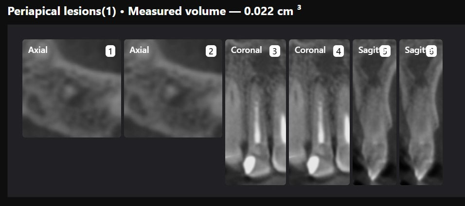
Measurement of periapical radiolucency volume is a unique advantage with 3D CBCT versus 2D PA. These are endodontic reports for tooth 12 which had a very large periapical radiolucency. The reports were done before and after a long-term calcium hydroxide dressing.
Third Molar Report
A single-third molar is selected to evaluate for extraction.
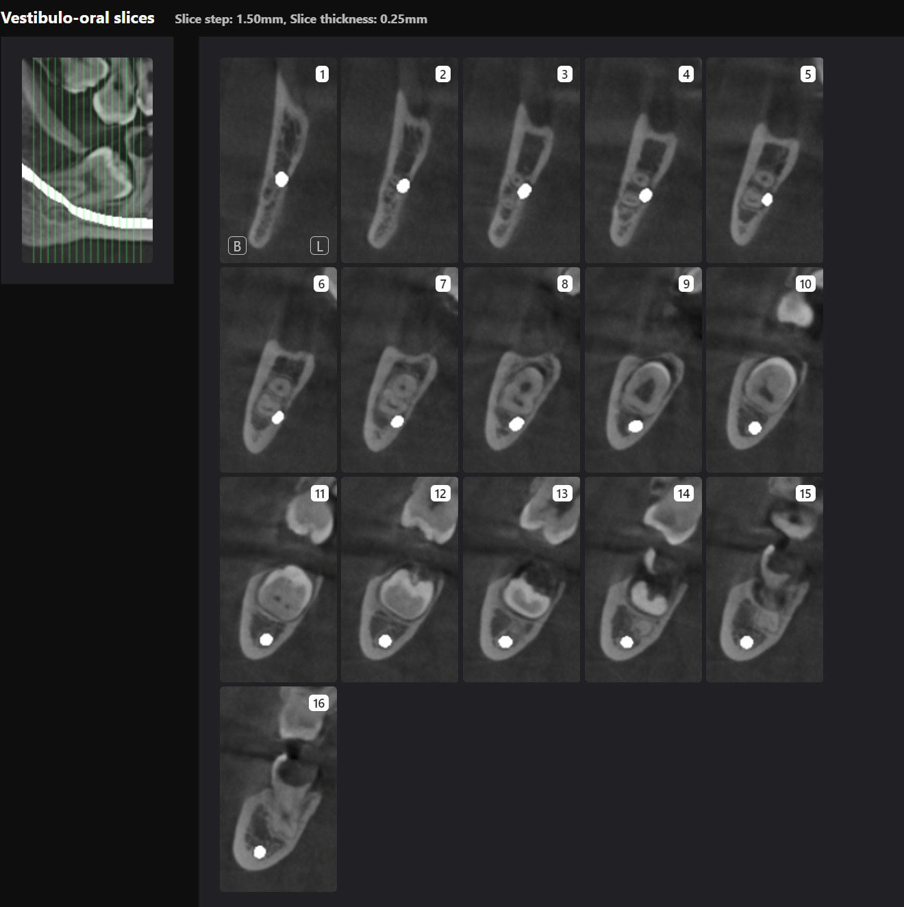
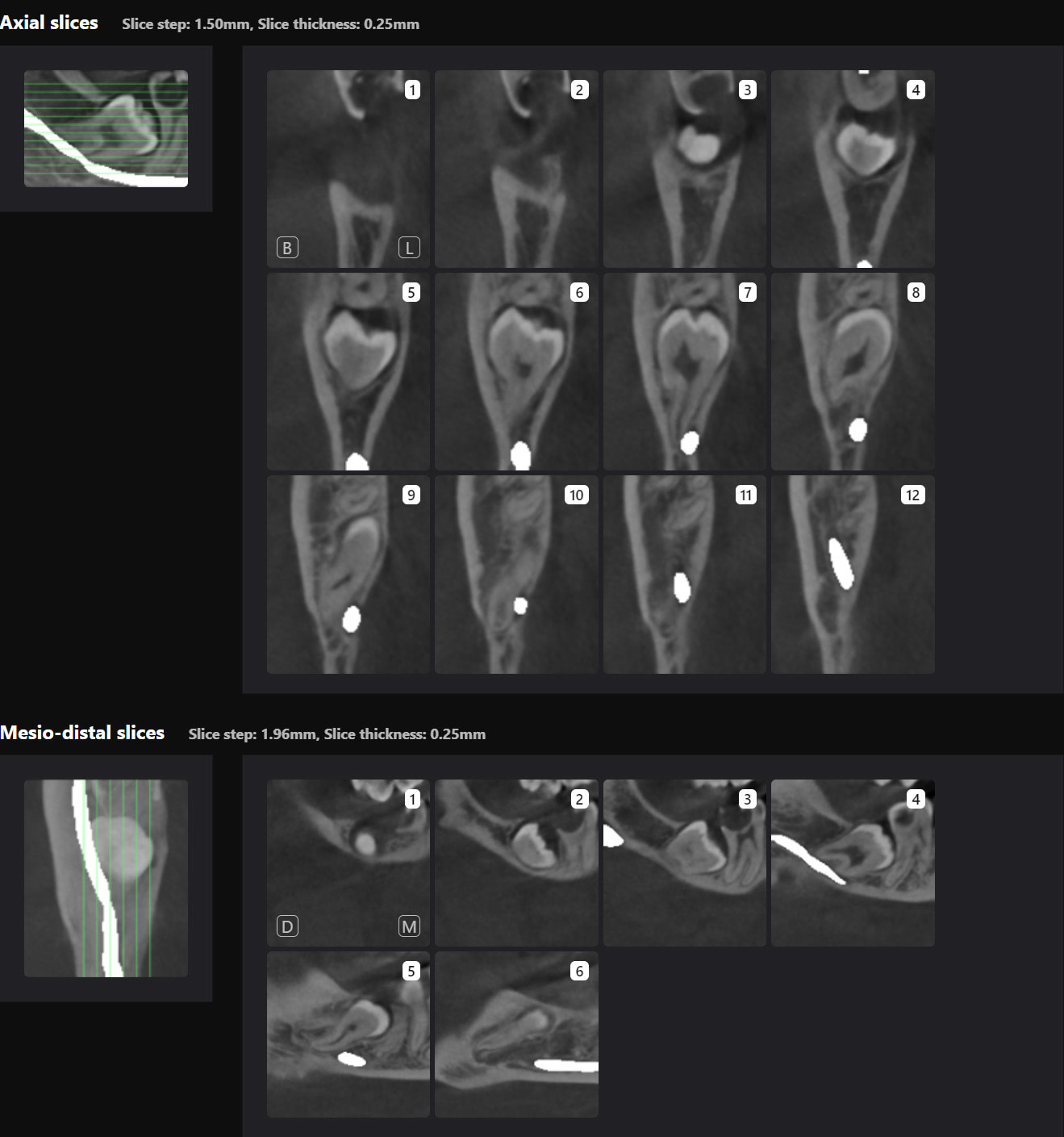
Generates slices in 3 directions and highlights the position of the inferior dental canal.
CBCT Segmentation into STL
Generate STL files from CBCT DICOM data.
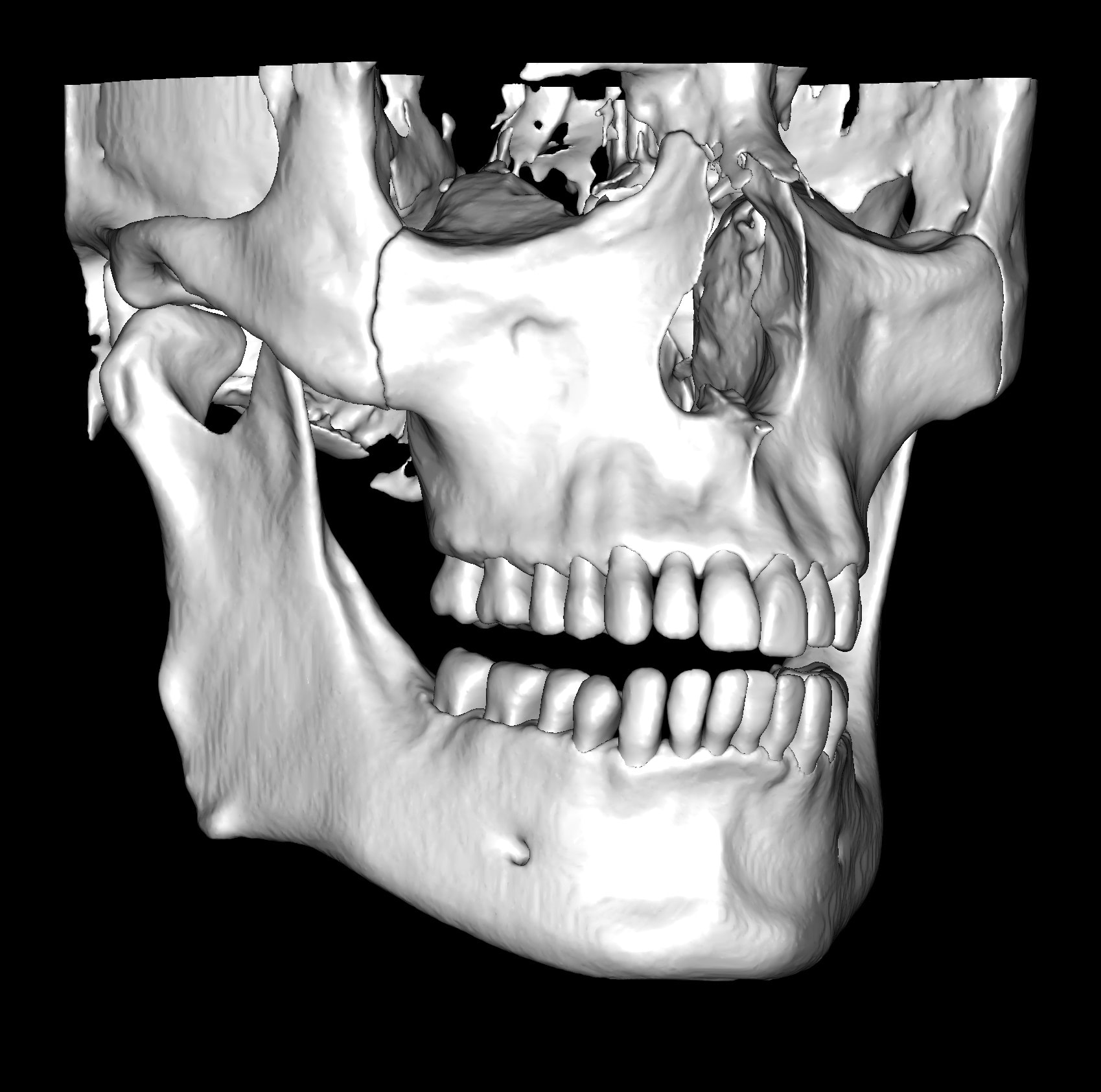
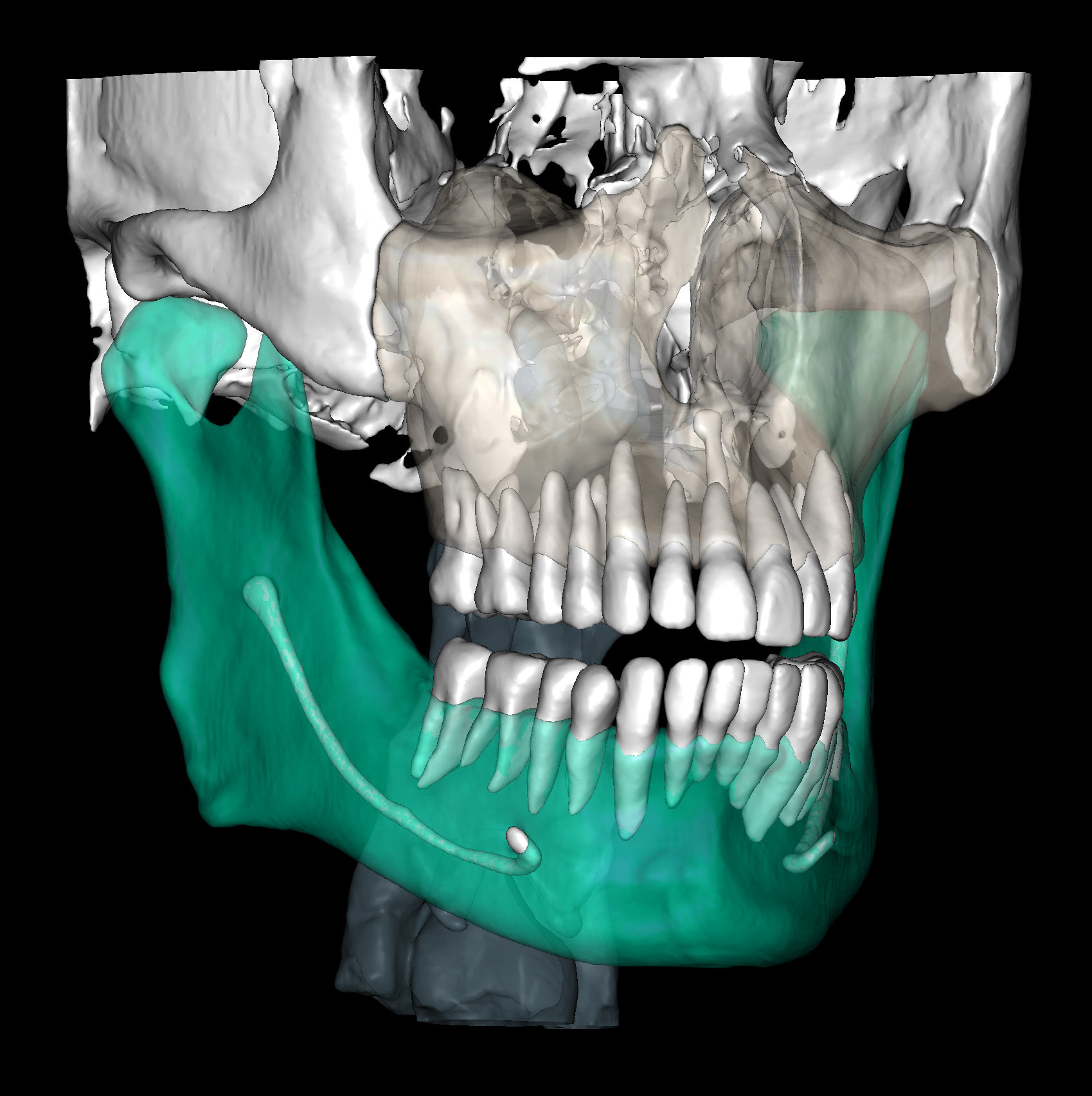
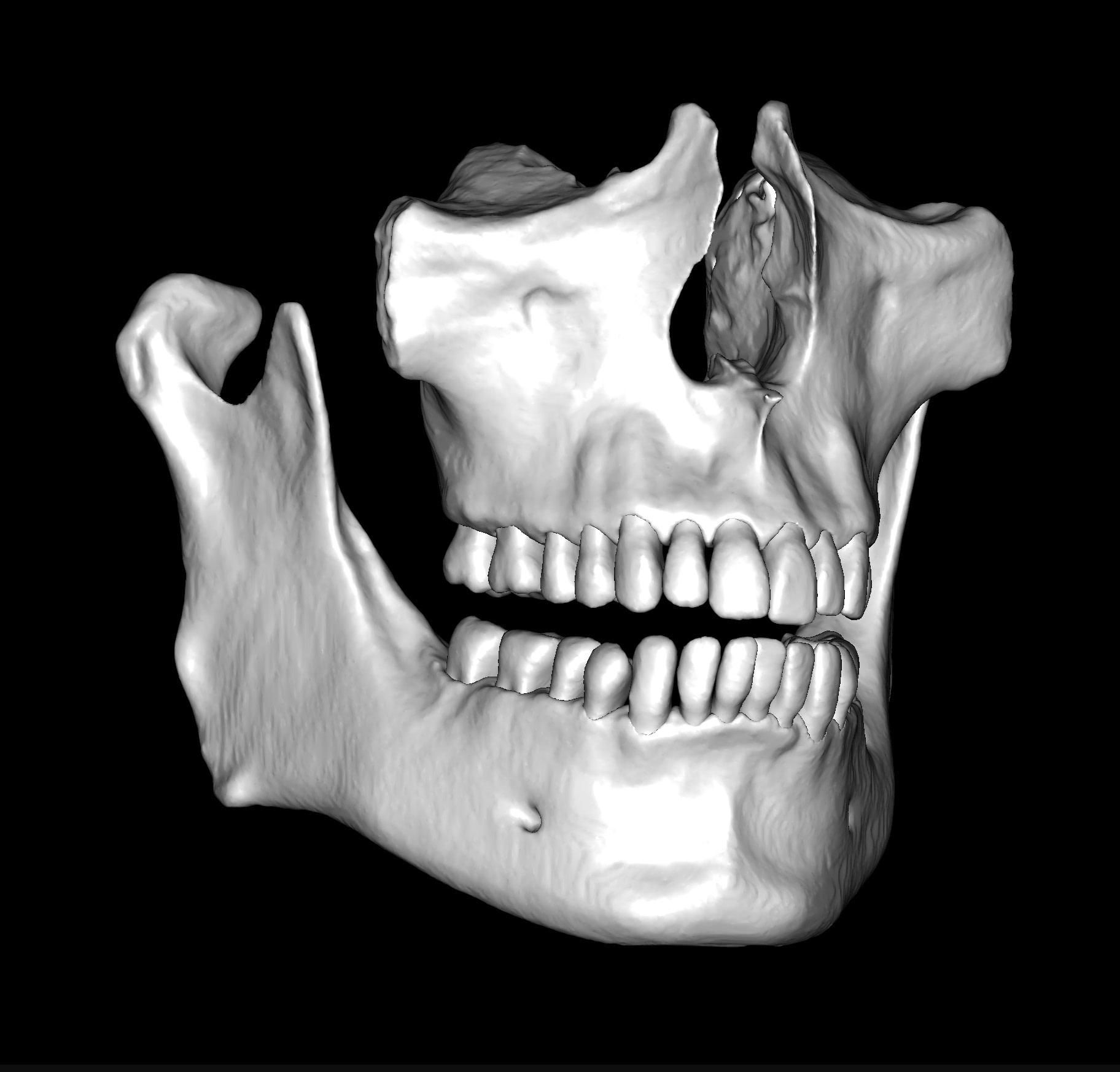
Generate maxilla and mandible in one STL file or face, teeth, maxilla, mandible, airway, cranial base, canals as separate STL files.
Creation of Digital Patient
The patient file in Diagnocat functions as cloud storage of photos, 2D and 3D radiographs, STLs, and documents.
Collaboration with colleagues and specialists
Thankfully, Diagnocat makes sharing easy. You can share the patient file with fellow Diagnocat users and colleagues who do not pay for Diagnocat services.
They can set up a Diagnocat account to view patient files shared with them. They can download the original files and view and download the Diagnocat reports. However, they are not able to order new reports.
How we use Diagnocat in our clinic
I used Diagnocat for all CBCT x-rays taken and most intraoral radiographs.
Typical Diagnocat workflow
- Take the radiographs at the beginning of the appointment
- Import radiographs into Diagnocat (automatically done with integration but can also be done manually).
- Order reports.
- Review radiological reports together with patients and approve findings.
- Sign and print the report.
- Upload a copy to the patient file (optionally, email it to the patient).
Importing the radiographs into Diagnocat is done one of two ways:
- Export the radiographs from your X-ray viewing software onto the desktop. Manually upload into Diagnocat patient file from there.
- Install the Diagnocat Desktop app:
- Export dicom files from normal viewing software into a designated folder on the desktop. The app will automatically upload files into the Diagnocat patient file using file data to identify the patient.
- Export directly from viewing software into Diagnocat patient file using the Export PACs function in viewing software.
How does Diagnocat compare to other AI diagnosis systems?
There are other dental AI diagnosis systems, such as Pearl and Overjet. We have yet to test these other systems at iDD. We will compare it to other systems in more detail in the future. However, when writing this article, Diagnocat is the only dental AI diagnosis system I have used to analyze 3D CBCT radiographs.
Areas for improvement
2D and CBCT reporting is excellent.
Orthodontic, endodontic, and implantology reports are basic, with room for useful extra details.
The orthodontic report generates OPG and front/lateral cephalograms. These are not as sharp as true OPG and cephalograms. Tracings of the maxilla, mandible, central incisors, canines, and molars are automatically produced on the generated frontal/lateral cephalogram.
I showed the orthodontic report to an orthodontist colleague. She found the tracings to be fairly accurate compared to a real lateral cephalogram of the same patient. She did further tracings on the generated lateral ceph and advised it was difficult to visualize points such as Nasion, ANS, A Point, Condylion, and Orbitae and visualize the fourth vertebrae to determine peak growth phase. Furthermore, other orthodontic software can automatically calculate skeletal/dental relationships, planes, and angles.
The company did inform us that they are working on full functionality sets for orthodontic reports. This will come next year.
The endodontic report could provide a better visualization of the shape of the root canals in 3 dimensions. It would be helpful to calculate the curvature angle to estimate the root canal's difficulty. Working length measurements of canals are shown automatically but measured from the pulpal floor. Typically, clinicians would estimate the working length of the radiograph by measuring from a common reference point such as the cusp tip or incisal edge.
The implantology report is especially basic and shows rudimentary ridge measurements in slices. There is no ability to plan a virtual implant (or restoration then implant for a restorative-driven workflow).
Again, the company is working on this. In fact, I did see a prototype they are working on that will automatically generate virtual implant placement and surgical guides - incredible stuff in the pipeline.
Occasionally, Diagnocat can make an erroneous finding. This is often due to artifacts on the 2D film phosphor plates, such as scratches/numbers or scattering on the 3D CBCT from movement/scattering from radiopaque objects such as metal restorations. The clinician must review all findings manually and confirm they are correct. Overall though, we were very happy and frankly, impressed.
We continue to use it in our clinic for our patients even after this review.
Future developments planned for Diagnocat
Alex (CEO) shared with us a bold vision of the future. Not only is the diagnosis done by artificial intelligence, but the treatment planning and treatment are completely automated!
He showed us the world’s first dental implant placement planning and surgical guide created completely by AI.
Also, they are planning clear aligner treatments that are completely automated and planned using CBCT, with STLs automatically generated to be used to produce aligners - all AI-driven. AI is so much more than just diagnosis and segmentation. We are excited for the future.
A caution regarding AI
AI can be used to guide medical diagnoses and treatment, therefore affecting real patients' lives and well-being. Current AI tools derive from machine learning or deep learning. Although we use the term AI, it is not a true human-level intelligence that is sentient and conscious.
It doesn’t necessarily follow the same logic as a human. Algorithms are derived by analyzing the data set that has been fed to it. It is not always possible to know how it came to decide on the finding. This is known as a black box model.
Any bias within the data set will reflect as a bias with the findings from the AI. For example, amalgam pins are widely used in New Zealand/Australia but are less common in other parts of the world. If the algorithm has never seen an amalgam pin before, it may give an incorrect finding that the tooth has been obturated with a root canal filling. I did initially notice this happening when we first started using Diagnocat around 12 months ago. However, with further updates, this appears to have been rectified.
It is an incredible diagnostic aid. Not the entire diagnostic process. Keep that in mind.
Download the Full Review PDF

Download the PDF to get a full copy
of this article to read later.
Get a high resolution printable copy of this review.
Conclusion
With the rapid advances in Artificial Intelligence and robotics, we are reaching a new era in human existence and healthcare. I believe we should approach this optimistically. It offers an opportunity to reduce the drudgery of day-to-day work and focus our concentration on critical thinking and making executive decisions. Artificial intelligence will still need our oversight and responsibility for its decisions.
I thoroughly enjoyed using Diagnocat and believe it is worth purchasing, particularly if you have a large clinic with CBCT.
I believe we are at the beginning of a flood of AI tools that will automate and speed up our work and reduce errors. One downside to this technology is that I completely changed my work using such tools for the better. But this makes it challenging to go back to working without it. It is similar to using intraoral scanners for years and returning to using physical impression materials.
Lastly, a final thought. I had an interesting comment from a patient - they were a bit uncomfortable to have AI diagnose them rather than a dentist who looked in their mouth. It is important to explain the role of AI to patients and that the dentist is ultimately making the diagnosis with the aid of AI.
Undoubtedly, we will continue to see the rise of this technology in dentistry. Not just diagnosis, but segmentation, data collection and analysis, treatment planning and even in CAD/CAM. Being a dentist has never been more exciting.

Hi I am Dr.Subhasish, from India, From where to get the diagnocat software.
Check the website and contact the company to see who is your regional distributor – https://diagnocat.com/en