iDD is back with another intraoral scanner (IOS) comparison. In this article, we compare several relatively affordable intraoral scanners, including IOS from Chinese manufacturers, with one of the most expensive scanners on the market.
At the Institute of Digital Dentistry, we are privileged to use all intraoral scanners within a clinical setting involving real cases and patients. This hands-on approach enables us to compare each intraoral scanner's performance objectively.
Dr. Ahmad Al-Hassiny scanned his patient, 14 crown, 15 inlay, 16 onlay, and 17 crown, with five different IOS on the same day. Click on the links below if you'd like to know more about each scanner through our unboxings and IOS reviews.

From left to right: Alliedstar AS 200E, Shining 3D Aoralscan 3, Runyes 3DS 3.0, 3Shape TRIOS 5 and CEREC Primescan.
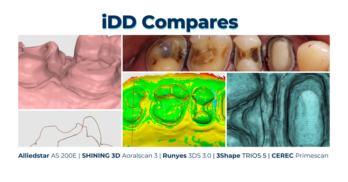
Here are the results of the individual scans - pictures of the color and monochrome scans, exported STLs, tessellated mesh, and a close-up of the prep margin to help you form your own opinion on these scanners.
Individual Scans in their Native Software
Every intraoral scanner available comes with its native scanning software. Most can eliminate scanning artifacts, such as movable soft tissues, cheeks, and tongue, using AI technology.
In the image below, you can see how these scanners capture color using their inherent software. How each scanner software adds ‘texture’ (color) varies and mainly depends on the scanner's precision in interpreting light reflection from the prepared area and surrounding teeth.

The processed color scans of the same site were captured using five different scanners, as previewed in their native software.
When looking at the color scans, we can see there are discrepancies in the scan brightness observed, such as the lower brightness of the 3Shape TRIOS 5 scan in contrast to the enhanced brightness of the Runyes 3DS 3.0 scan, which we’ve previously observed in other iDD compares cases.
The saturation and brightness of the scans captured using the Alliedstar AS 200E, Shining 3D Aoralscan 3, and CEREC Primescan IOS appear to be similar, with the Alliedstar AS 200E possessing slightly more surface texture within the color scan as previewed in its native scanner software.
Some scans look cartoony, some look realistic, and then there is the ‘hyper-realistic’ look with high-definition appearances. What is your favorite?
Moving on, let's compare the monochrome scans.

The processed monochromatic scans of the same site were captured using five different scanners, as previewed in their native software.
Monochromatic scans can also be taken and previewed in their native software. These scans provide a better view of the quality of the prep, and it is recommended that you use monochrome filtering to check for any scan issues that would be less obvious when viewed in color.
Here, you can see some apparent differences between the scans. Some margin lines (notably TRIOS 5 and Primescan) are prominent, while others blend slightly with the surrounding soft tissue.
Exported Scans in Third-Party Software
All intraoral scanners have an open architecture permitting scan exports. These files are exported in three formats: STL, PLY, or OBJ. All scanners can export STL, but not all do PLY and OBJ. Notably, CEREC only exports in STL.
While STL files are monochrome scans, OBJ and PLY files can capture and store color and texture details.
The scanners used in this iDD comparison are capable of exporting scans in the following formats:
- Shining 3D Aoralscan 3 and Runyes 3DS 3.0 - STL, PLY, and OBJ
- Alliedstar AS200E and 3Shape TRIOS 5 - STL and PLY
- CEREC Primescan - STL only
STL File Sizes Compared
Alliedstar AS200E
Aoralscan 3
Runyes 3DS 3.0
3Shape TRIOS 5
CEREC Primescan
PLY File Sizes Compared
Alliedstar AS200E
Aoralscan 3
Runyes 3DS 3.0
3Shape TRIOS 5
CEREC Primescan
OBJ File Sizes Compared
Alliedstar AS200E
Aoralscan 3
Runyes 3DS 3.0
3Shape TRIOS 5
CEREC Primescan
Based on the sizes above, the scans captured by the Alliedstar AS200E scanner are the largest.
Dental labs often utilize independent CAD software. In our case comparison, we used Medit Design to view the exported STL, PLY, or OBJ files and design restorations based on these scans. This approach allows for an impartial viewing of the scans, independent of the individual scanner's native software, thereby bypassing any customized color or optimized surface rendering provided by the proprietary software.

All scans were exported in an STL format and previewed in the Medit Design app.
Scan Accuracy
Using Medit Design, we looked closer at the amount and detail of data captured within each scan.
When we observe the close-up of the preps and their tessellated mesh, the scan captured using the Alliedstar AS200E appears to have the densest mesh. Referring back to the exported file sizes table mentioned in the previous section, the mesh density supports the scan's large STL (and consequently the PLY) file size.
Click on any image to view at full size.
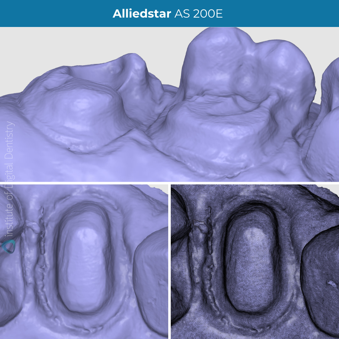

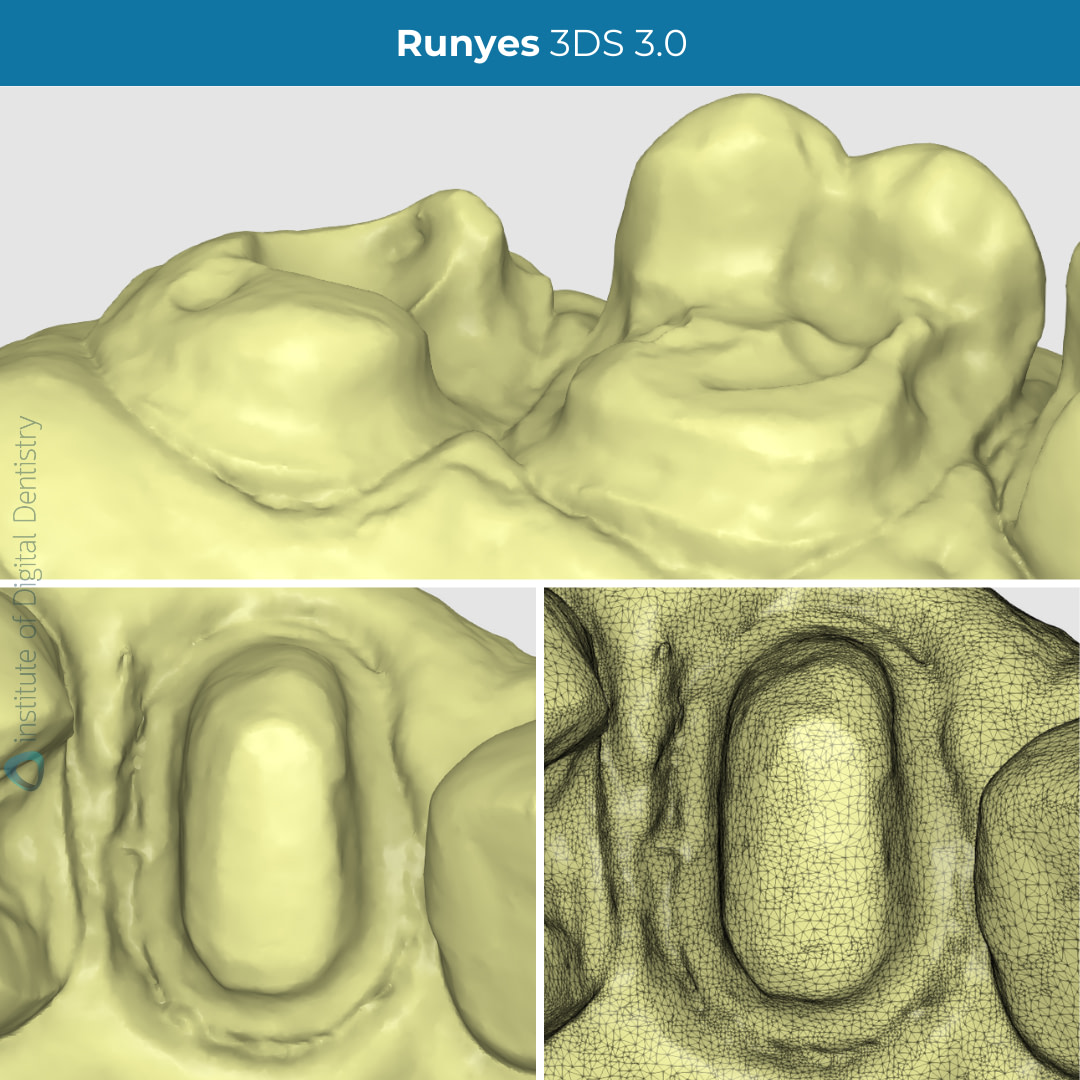
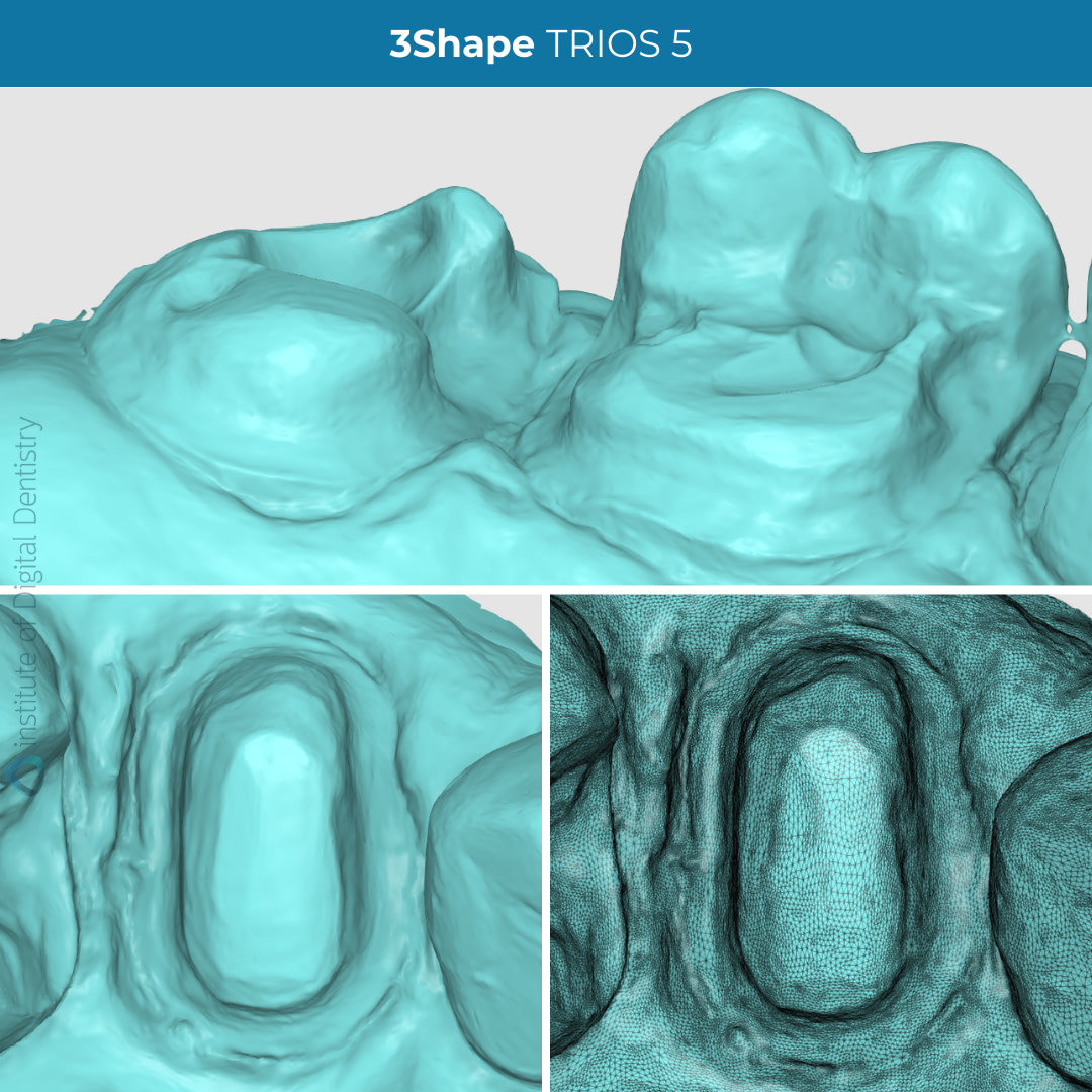
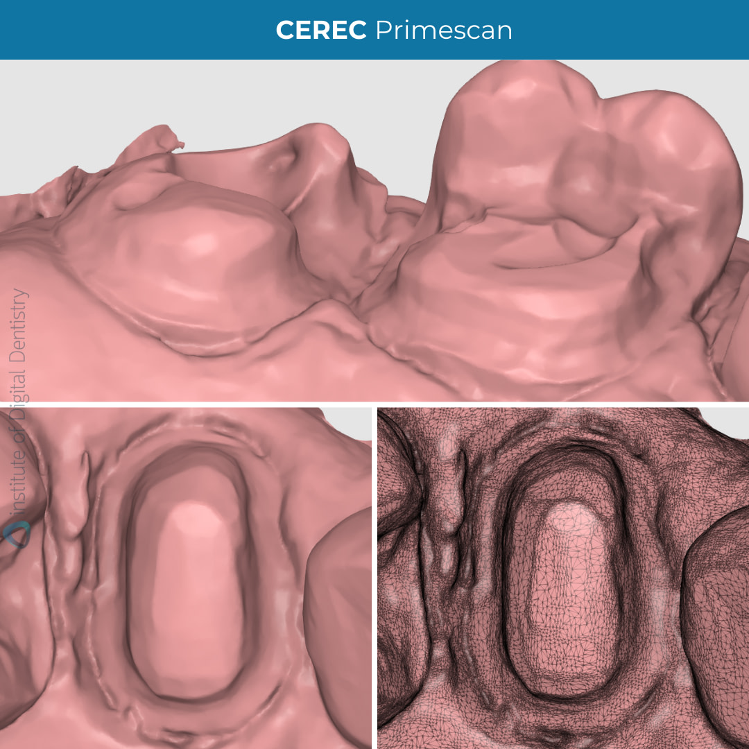
When we observe the close-up of the preps and their tessellated mesh (16-17 mesio-palatal prep margin line, 14 crown prep, and tessellated meshes of each scan as previewed in the Medit Design app), the scan captured using the Alliedstar AS200E appears to have the densest mesh. Referring back to the exported file sizes table mentioned in the previous section, the mesh density supports the scan's large STL (and consequently the PLY) file size.
3Shape TRIOS 5 follows closely behind AS200E with a similarly dense mesh. With this being said, Shining 3D Aoralscan 3, CEREC Primescan, and Runyes 3DS 3.0 scans are also dense. There are yet to be any studies investigating the clinical significance of mesh density. Therefore, a denser mesh does not necessarily indicate a ‘better scan.’ Currently, we can view the relationship between how the scan file size correlates with the scan’s mesh density.
Medit Design or third-party CAD software can also review prep margin lines. Modern intraoral IOS devices operate by projecting light onto the surfaces to be scanned. This light must be reflected to the IOS from the scanned surface for a precise scan. If the light penetrates the surface without reflecting, this leads to inaccurate scans.
Generally, IOS devices may encounter challenges with equi or subgingival preparations (though this can be mitigated with appropriate tissue and moisture management). Although Primescan is known to be one of the most accurate IOS on the market, the TRIOS 5 appeared to have captured the most detailed, prominent, visible margin line. 3Shape scanners are regarded as quality and accurate devices, with studies backing these claims.

Deviation map of the scans compared to CEREC Primescan scan and sectional view. There seems to be very little deviation around the prep area.
CEREC Primescan was allocated as our point of reference in which we can view the deviations of the scans when aligned using Medit Design’s Deviation Display mode. Based on the colored deviation key, we can see that the scan meshes are -0.025 to +0.025 mm deviation compared to the Primescan reference.
There seems to be some slight deviation between the different scanners, in particular in the areas around the 17 mesial margin prep line, 16 buccal surfaces, 15 inlay prep, and 14 distal prep surface and margin line, which exhibit a deviation of around +0.025 mm or 25 microns. Is 25 microns clinically significant? Likely not when the cement gap itself is 70-120 microns
Within the same Deviation Display mode, we can view the aligned scans in a sectional view in which we can see minimal differences between the scans.
Conclusion
While minor variations are noticeable across the five scans, either within their native software or third-party applications, no substantial deviation exists between all scans and the ability of these scanners to capture these preparations.
The accuracy in capturing the edge of the preparation area might slightly differ depending on the individual scanner, which could affect the dentists' or technicians' margination. Soft tissue retraction is, as always, critical
It is still not clear if differences in scanner software and hardware significantly affect the accuracy of digital impressions from different companies. More research is needed in a controlled clinical setting to reach a more definitive conclusion about the many scanners available. The iDD team is investigating this.
Finally, the final restorations are milled in-house using CEREC Primescan and Primemill and completed in a single visit.

Which IOS or type of cases would you like to feature in an iDD Comparison?
Leave a comment below about what you’d like us to cover next.



Panda Smart and a cross arch accuracy study to test the significance of photogrametry would be a blow
We carried out tests to evaluate the cross-arch accuracy of photogrammetry systems like the Panda Smart. Preliminary results indicate that while these systems offer significant advantages in workflow efficiency, premium intraoral scanners still seem to have a slight edge in full-arch implant accuracy. We’ll share more comprehensive findings as the studies progress.