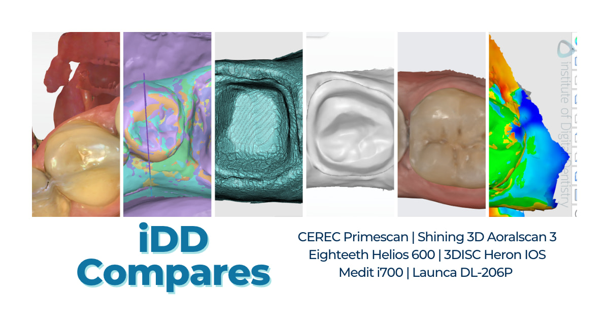
I scanned the same patient, on the same day, with six different intraoral scanners:
- CEREC Primescan by Dentsply Sirona
- Aoralscan 3 by Shining 3D
- Heron IOS by 3DISC
- Helios 600 by Eighteeth
- i700 by Medit
- DL-206P by Launca
All these scanners, with the exception of CEREC Primescan, have their recommended retail price between $9-18k USD.
Yes, even Medit i700 whose price has been reduced due to the release of the i700 Wireless.
At the Institute of Digital Dentistry we are very fortunate to be in a position to test all intraoral scanners in a clinical environment, on real cases and real patients.
Below I will show you the pictures of the color scan, exported STLs, and the tessellated mesh to help you form your own opinion on these scanners.
Scans in their Native Software
Every scanner comes with its own scanning software.
Simply put, if the scanner is what’s capturing the surface, the software is stitching it up together. Modern intraoral scanners come with AI that is responsible for removing various scan artifacts (e.g. movable soft tissue, cheeks, and tongue) to various levels of success.
First, let’s have a look at how these scanners capture color and how they preview it in their native software, which becomes handy when marking equi- or subgingival finishing line.
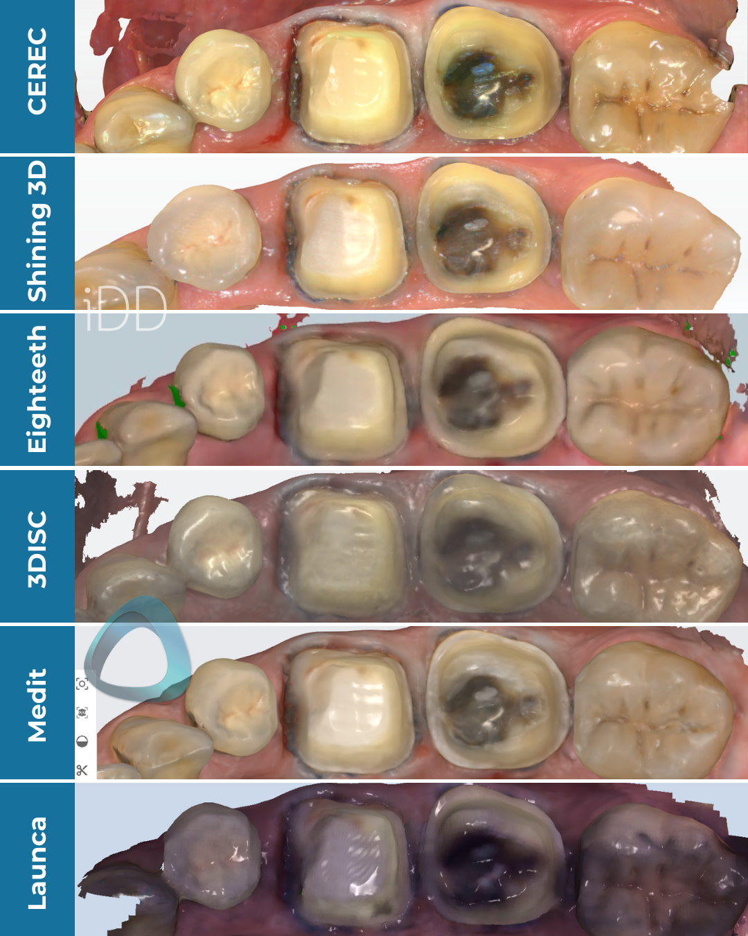
The same tooth preparation captured with six different scanners, processed color scans as previewed in their native software.
As you can see, every scanner captures color slightly differently, with the biggest difference being observed in the scan taken with the Launca DL-206P.
In the next picture, the scans are still previewed in their native software, but in monochrome.
I would recommend checking your scans in this mode from time to time as it makes stitching issues, holes, over-scanning, and other issues more obvious.
This view also gives you a better idea about the preparation quality and roughness as well.
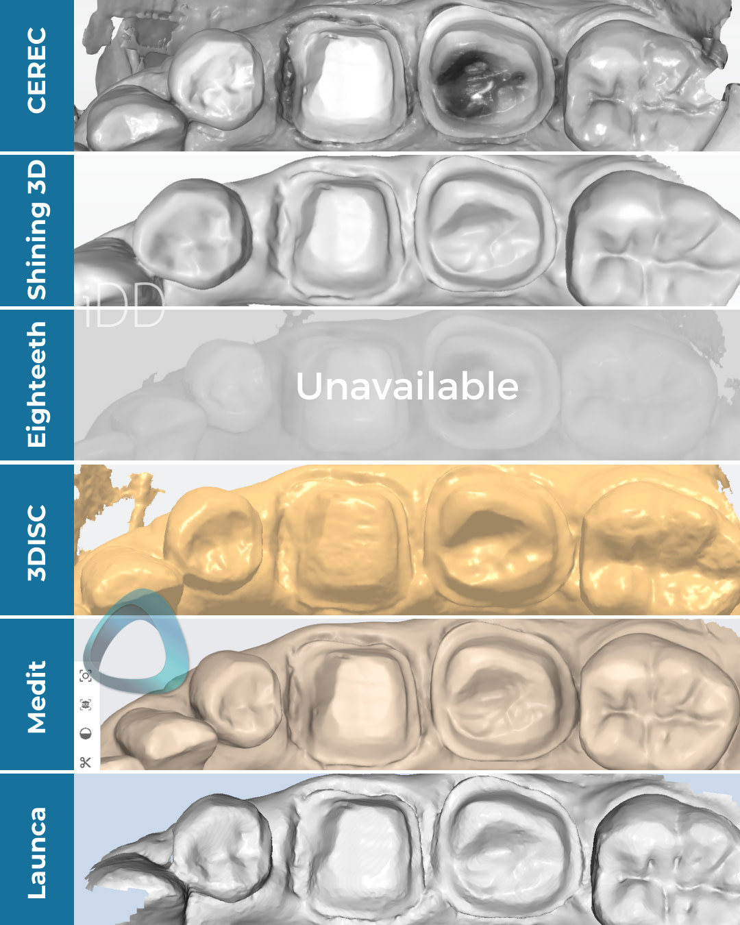
Scan meshes in monochrome previewed by their native software. Unfortunately, we were unable to capture Helios 600’s mesh due to the system’s default settings.
Exported Scans in Third-Party Software
The next series of pictures shows the exported scans in the Medit Design app.
Intraoral scanners nowadays have an open architecture that allows you to export the files and send them to a lab.
These scans can be exported in various formats, usually monochrome STLs, and PLY or OBJ files that also carry the color information.
In this particular set of scanners, the ones capable of exporting scans in these formats include:
- i700, Heron IOS, Aoralscan 3 - STL, PLY and OBJ
- Helios, Launca - STL and PLY
- Primescan - STL only
That being said, we exported scans in an STL format in their default settings.
The Medit i700 and Aoralscan 3 scans were NOT captured in HD.
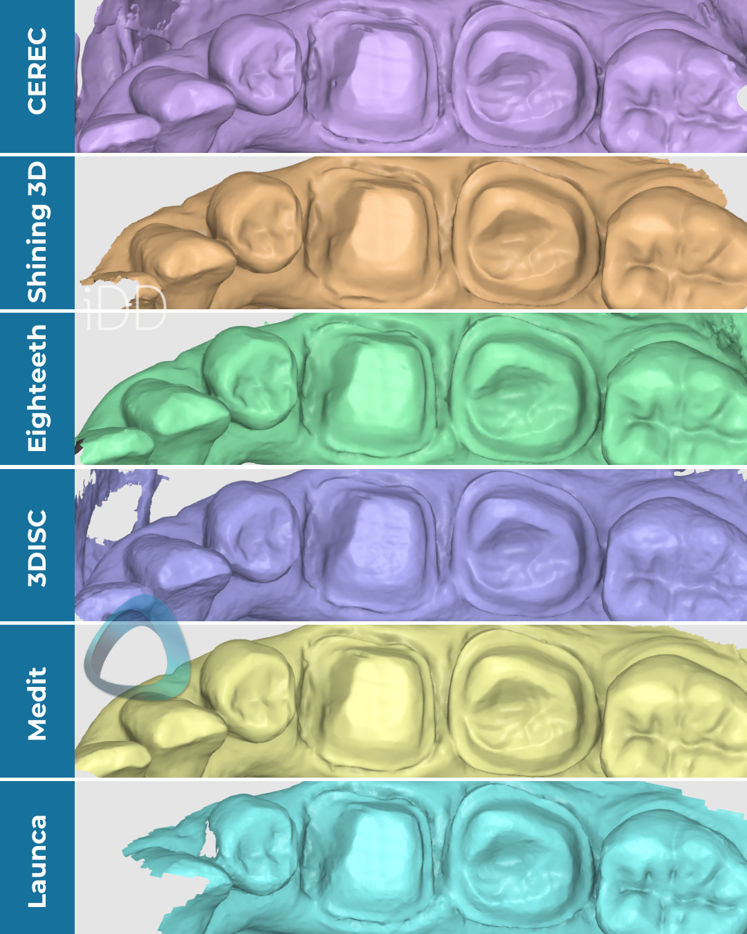
All scans were exported in an STL format and previewed in the Medit Design app.
The goal of this is to prevent scanners from hiding behind their custom color and texture rendering.
Most of these scanners are considered to be impression-replacement tools for dentists who plan to send the scans to dental laboratories. Dental technicians in these labs will then create restorations using third-party CAD software.
Therefore, it’s crucial to see the scans outside their native software, where they can't hide behind fancy surface rendering.
In the next picture, you can also see the close-up of the preps and their tessellated mesh.
This reveals the amount and detail of data captured.
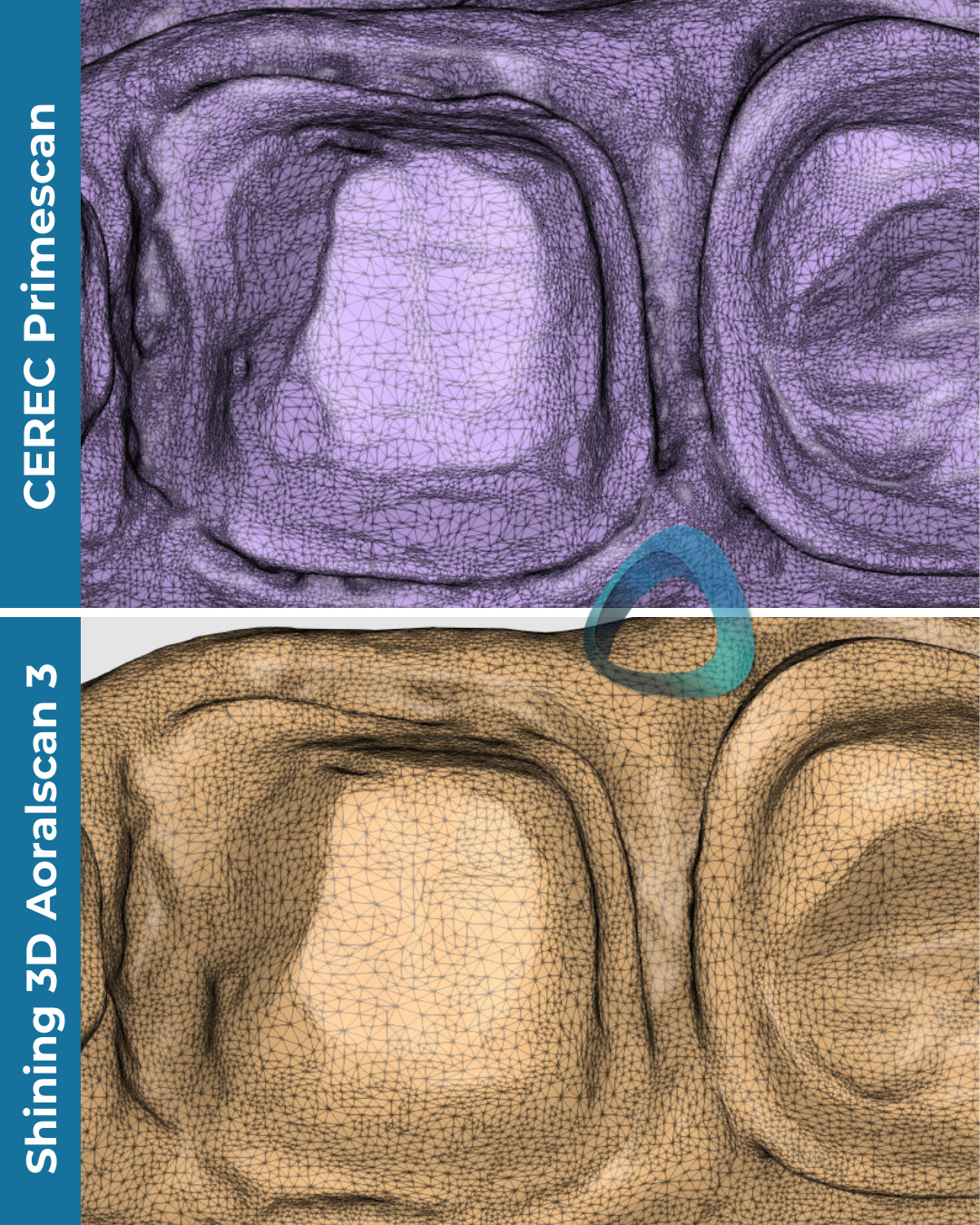
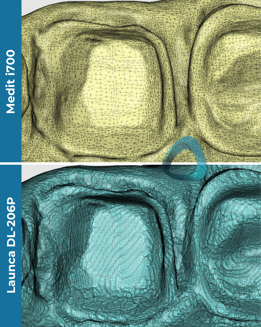
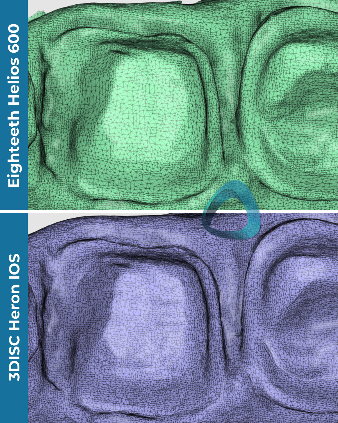
Primescan and Heron IOS seem to have the densest meshes, which presumably means capturing the most details but not always.
On the other hand, Launca DL-206P’s mesh also seems to be quite dense but in this case, a denser mesh doesn’t necessarily translate into a ‘better scan’.
Unfortunately, no studies from my understanding have been done regarding the mesh density in relation to clinical significance.
Scan Accuracy
The accuracy and trueness of most of these scanners is not backed by any independent research, the only exception being CEREC Primescan. Primescan is commonly regarded as the most accurate scanner, which is why we chose it as a reference.
First, we aligned all the meshes. Using the Medit’s Deviation Display Mode, it was clear that these meshes were all fairly similar compared to the scan taken with CEREC Primescan.
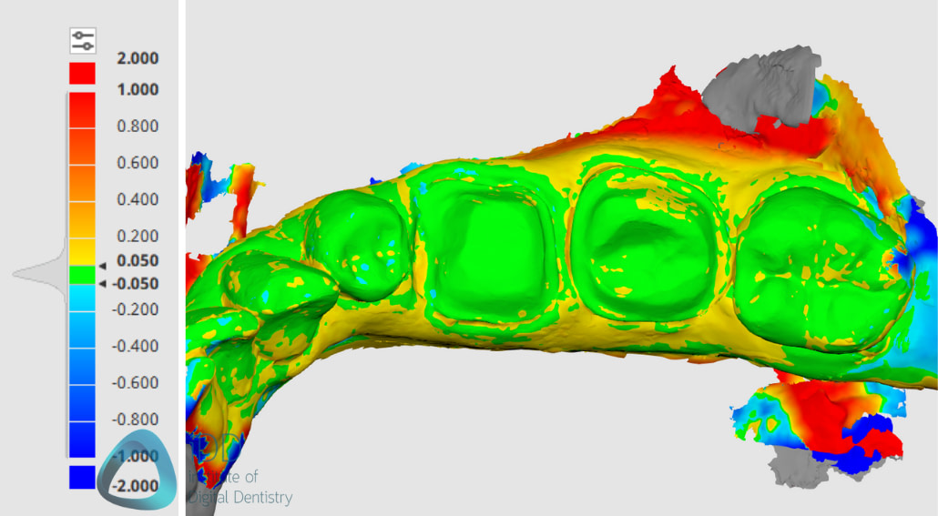
Deviation map of the scans as compared to CEREC’s scan. There seems to be very little deviation around the prep area.
In the next picture, you can see the same aligned scans but this time in a sectional view.
It again looks like there is very little difference between the scans.
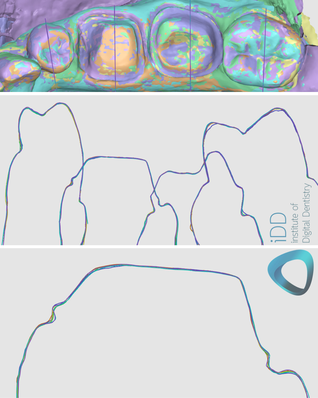
The sectional view of the over-imposed scans.
Conclusion
There is a noticeable difference between these scans, both within their native systems or third-party software but we did not observe a significant deviation of one scan from another in terms of accuracy.
However, the way these scanners capture the edge of the preparation might affect subsequent margination by dentists or technicians.
From this case alone, it’s not conclusive whether there is any clinical significance in the different ways software produces digital impressions.
More in-depth research in a controlled environment is definitely necessary to draw a proper conclusion about the wide range of scanners on the market. In our experience, we have used all these scanners in the practice with no issue clinically for simple things like crown and bridge work.
What are your thoughts? Let us know in the comments below!

Hi dr, big tnx for the great info????
And also would appreciate if you explain the margin accuracy comparison results between Launca DL-300 and other ios devices?
Thank you so much for your kind message, really appreciate it! I’m glad you found the info helpful.
Once i get access to a DL-300 I will put together something for sure.