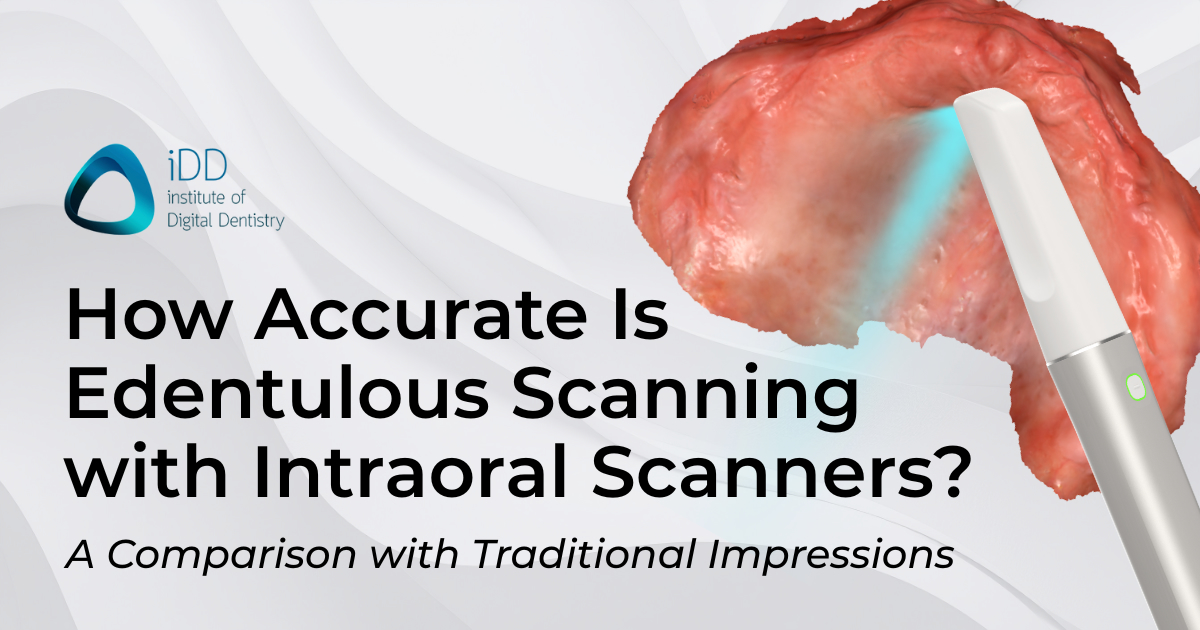
At iDD, we often get asked by clinicians and technicians alike, Is edentulous scanning possible with an intraoral scanner (IOS), and is it accurate?
The short answer is yes. But it's not always easy, and you need a great scanning technique.
Additionally, edentulous scanning is the one indication that totally differentiates the various IOS brands. While a single crown prep can be scanned with basically any scanner these days, some IOS are far better for edentulous scanning compared to others.
Edentulous scanning has long been considered the final frontier of digital impressions. For years, many dentists believed it was impossible to accurately capture a fully edentulous arch with an intraoral scanner. And let's be honest, they weren't entirely wrong. After all, a muco-compressive impression is not the same as a digital impression, which is muco-static.
But here's the thing, technology doesn't stand still. Today's intraoral scanners differ significantly from those of a few years ago. Companies like 3Shape and Medit, as well as more recently SHINING 3D with the release of their Aoralscan Elite IOS, have been pushing the boundaries of what's possible with digital scanning.
Also, our understanding of CAD and designing dentures for muco-static impressions, such as digital scans, has improved significantly, leading to retentive dentures even when using just an IOS.
Despite the challenges and the learning curve, I firmly believe that digital scanning for edentulous cases is 100% possible and can even be better than traditional methods. So, let's break down why.

The End of Distorted Impressions
We hire 6 technicians at iDD Lab. Many started their careers using totally traditional techniques. They can't count the number of times they have opened a case box to find distorted or inadequate physical impressions. This is a sentiment many technicians can relate to.
Each one meant hours of extra work or, worse, asking the dentist for a retake. It was frustrating for everyone involved - the lab, the dentist, and, most importantly, the patient. Digital scanning changes all of this. Yes, it has its challenges, but the benefits are huge.

So let's get into these challenges that some would argue against edentulous intraoral scanning:
- No fixed landmarks - when scanning a dentate arch, teeth serve as reference points. With an edentulous arch, these fixed points are absent, making the stitching process more challenging.
- Mobile soft tissues - including the cheeks, tongue, floor of the mouth, and even the soft palate, can all move during scanning, potentially leading to stitching errors, inaccurate scans, double images, and a frustrating process.
- Large scanning area - an edentulous scan typically requires capturing a much larger area than a single crown or even a full-arch scan with teeth - maxillary tuberosities, retromolar pad area, etc. (hint: large FOV scanners make this easier).
- Undercuts - capturing undercuts is absolutely necessary for denture retention, but can be tricky for scanners, especially for newbies.
- Saliva management - edentulous patients can be more challenging to manage, not only due to soft tissue issues but also due to excessive saliva. Especially lower arch scanning. Pooling of saliva = poor scan quality.
In the face of all that, with digital scanning, you can immediately see if you've captured everything you need. You don’t need to wait hours or days to find out from your technician if your impression meets the standard, and you can rectify any mistakes almost immediately.

Modern Intraoral Scanners are Game Changers for Edentulous Scanning
The latest generation of intraoral scanners has a number of features to help with edentulous scanning:
- Larger fields of view - scanners like the iTero Lumina, Medit i900, and Aoralscan Elite all have large scan head FOVs. This appears to be a current market trend. This means they can capture more in a single image, which typically results in a faster scan and fewer stitching issues.
- Improved stitching algorithms - AI-powered software is becoming more adept at assembling those challenging edentulous scans. For example, 3Shape ScanAssist for the TRIOS 5 and 6 is particularly good at reducing stitching problems. In fact, TRIOS scanners have been excellent at edentulous scanning since TRIOS 3.
- Better soft tissue scanning and specific edentulous modes - many modern scanners can now capture soft tissue detail much more effectively and have toggles for edentulous scanning - this fine-tunes the software AI for the different demands of capturing soft tissue.
- Improved scanning speed - faster scanning means less time for soft tissue movement or saliva accumulation.

The ability to receive detailed, accurate digital scans of edentulous arches eliminates many of the variables we used to deal with, like impression material distortion or model pouring errors. In the end, though, good scanning technique is paramount - especially good soft tissue retraction. Technology can only do so much for us.
Precision That Traditional Methods Can't Match
Now, I know what some of you are thinking. "But traditional impressions can capture every detail perfectly!" And you're not wrong - in theory. A perfectly taken traditional impression with a high-quality material can capture incredible detail.
But let's be real: how often does that actually happen? In the real world, with real patients who move and produce saliva, and real dentists who are pressed for time, traditional impressions often fall short.
Digital scanning enables a level of precision that is nearly as good and consistently hard to match with analog techniques. The literature has proven this countless times.
“The clinical performance of restorations fabricated based on intraoral scans has proven to be comparable to those obtained using conventional impressions, substantiating the reliability of IOSs in restorative dentistry.” Read it here.

The Secret is Scanning Technique
The secret is all in the technique and practice.
It’s often the downfall of clinicians, dentists, and dental technicians alike, who argue against using an intraoral scan with their edentulous scan because they lack the correct technique or are unwilling to adopt such a technique.
Here's something we always emphasize at iDD: even the best technology needs good technique. When it comes to edentulous scanning, the technique is crucial and remains crucial for a scan to succeed.
Here are some tips:
- Retraction is key: Use cheek retractors / a mirror and have your assistant help manage soft tissues. For the upper arch, you need to retract the lips at all times and retract them so well that the sulcus and movable tissue are immobile. The same is true for the lower arch, but you also have to deal with the floor of the mouth and the tongue.
- Note: There are countless retractors on the market now. Use whatever you like. I have not found one particularly better than the other. I personally just use my finger for the upper and a mirror for the lower.
- Dry the tissues: It depends on the patient. Some have mostly dry mouths, and you are fine scanning. Others, it is a pool of saliva, especially lower scans. Passing a suction tube around the mouth can help reduce reflections and improve scan quality.
- Strategic scanning: know the correct scan strategy for edentulous scanning and where you are going. Typically, you should start on an area that does not move and has good reference points, such as rugae, residual ridge, retromolar pad, etc. Then work around the arch, the vestibules, and finally, the buccal and lingual sulcus.
- Take your time: Edentulous scanning isn't a race. Slow and steady wins here. That being said, scanning the upper and lower arches doesn't take 15 minutes, but it can take around 2-3 minutes per arch to do properly.
- Review as you go: Use the software's real-time review feature to identify areas that need rescanning or may need to be deleted and repeated. Watch the screen while scanning, not the patient.
- Note: If you encounter a significant stitching error, as painful as it is, it is often easiest and best to simply delete the entire scan and start again. Rescanning the area sometimes fixes the issue, but often not.
I want to emphasize the importance of good technique. Even the best scanner can't compensate for poor retraction or a rushed job.

The Case for Traditional Impressions
While we're excited about the advancements in digital scanning, we can't ignore the fact that traditional impressions still have their place. Here's why some clinicians are sticking with the analog approach:
- Predictability: Let's face it, traditional impressions are the devil we know. Many dentists have years, if not decades, of experience with them. They know exactly what to expect and how to handle any curveballs that come their way.
- Material advantages: Some argue that impression materials can capture undercuts and soft tissue details more accurately than current scanners. There's something to be said for the way a good polyvinyl siloxane impression can flow into every nook and cranny and compress the tissues.
- No technology barriers: Traditional impressions don't require expensive equipment or software updates.
- Skill transfer: Many clinicians have spent years perfecting their impression technique. Starting from scratch with digital methods can be a tough pill to swallow.
The Learning Curve is Worth It
Now, I'm not going to sugarcoat it; there is a learning curve with digital scanning, especially for edentulous cases. It's not just about learning to use the scanner; it's about rethinking your entire approach to case planning and execution.
But here is the undeniable truth - IT IS TOTALLY WORTH IT.
The speed, precision, and predictability you gain, especially for all cases, are invaluable. And let's not forget the "wow factor" for patients. Or the fact that patients prefer having a scan done rather than an impression. With intraoral scanners, you never have to worry about a gagging patient or asking them, “How are you with impressions?”.
In my experience, patients are consistently impressed by digital technology, which can lead to higher acceptance rates for cases. We literally have patients coming to some of our denture clinics we run in New Zealand saying, "I heard you guys are the ones who use the scanners". It's a practice builder.
Cost Considerations: Think Long-Term
Yes, intraoral scanners are a significant investment, often ranging from $3,000 to $20,000 or more.
But in my view, it's short-sighted to focus solely on the initial cost.
Consider this:
- Long-term savings: Digital workflows can lead to savings on impression materials and lab fees over time.
- Increased efficiency: Potential for increased productivity and faster turnaround times.
- ROI timeline: The time to see a return on investment varies depending on practice volume and type.
- Increased case acceptance: Many clinicians report higher acceptance rates with digital scans.
- Don't forget this key detail - many labs around the world are now refusing to accept physical impressions. I am not joking. Times are changing.
Clinical Outcomes
At the end of the day, what matters most is the outcome for our patients.
So, how do digital and traditional methods compare?
- Accuracy comparisons: Studies have shown that digital scans can be as accurate, if not more accurate, than traditional impressions for many applications, even for complex edentulous cases. A 2019 study in the Journal of Prosthetic Dentistry found comparable accuracy between digital and conventional methods for complete dentures but noted that more research is needed.
- The fit of final prosthetics: In our experience, digitally designed dentures can fit just as well as traditional ones - sometimes even better. However, it depends on the case and the skill of both the clinician and the lab. It is all in the scan, design, and manufacturing prowess. It's all in the CAD.
- Patient satisfaction: This is where digital often shines. Many patients prefer the comfort and speed of digital scanning over traditional impressions. Plus, the "wow factor" of seeing their scan on screen can be a great practice builder. Don't forget the fast turnaround time for digital dentures. 24-hour dentures are a thing now.
- Integration with other digital workflows: Digital scans can be easily integrated with CAD/CAM milling or 3D printing. This results in a much faster workflow. Setting up denture teeth digitally is 10 times faster than doing it physically. It's not even comparable.


The Future is Digital
As someone who's worked on both sides of dental technology, using traditional and digital methods, I'm convinced that the future of edentulous case management is digital. It is time to accept that this is the standard of care and start learning how to do it.
Here's what I expect to see in the future:
- Continued technological advancements: Even better soft tissue capture and more advanced AI for processing scans.
- Industry-wide digital shift: More labs and manufacturers are moving towards digital workflows.
- Education evolution: digital dentistry will not only be introduced but also implemented as part of dental education for future generations.
- Better materials: especially in the 3D printing space. 3D-printed dentures are improving at an incredible rate and will soon be an industry-wide accepted alternative to traditional denture manufacturing. Milling dentures is still king, thoug,h at the moment, for long-term dentures 🙂
Conclusion
So, is edentulous scanning possible? Absolutely. Is it easy? Not always. But with training and practice, yes.
Digital scanning for edentulous cases is becoming increasingly viable, but it's not a magic bullet. It requires investment in equipment, training, and patience as you climb the learning curve. Going digital is undeniably the way forward. The benefits in terms of accuracy, efficiency, and patient experience far outweigh the initial challenges.
To my fellow dental professionals who are undecided about digital scanning for edentulous cases, I say this: take the plunge. Invest the time to learn the technology and refine your technique. Your future self - and your patients - will thank you.
Digital scanning for edentulous cases isn't just a trend; it's the future of dentistry. And in my opinion, that future is bright.
My advice? Stay informed, keep an open mind, and be willing to adapt as technology evolves.
Whether you're in team digital, team analog, or somewhere in between, the goal is always the same: to provide the best possible care for our patients.
What's your experience with edentulous scanning? Whether you're a dentist or a technician, we'd love to hear your thoughts in the comments below!
And as always, we at iDD are here to support you on your digital dentistry journey.