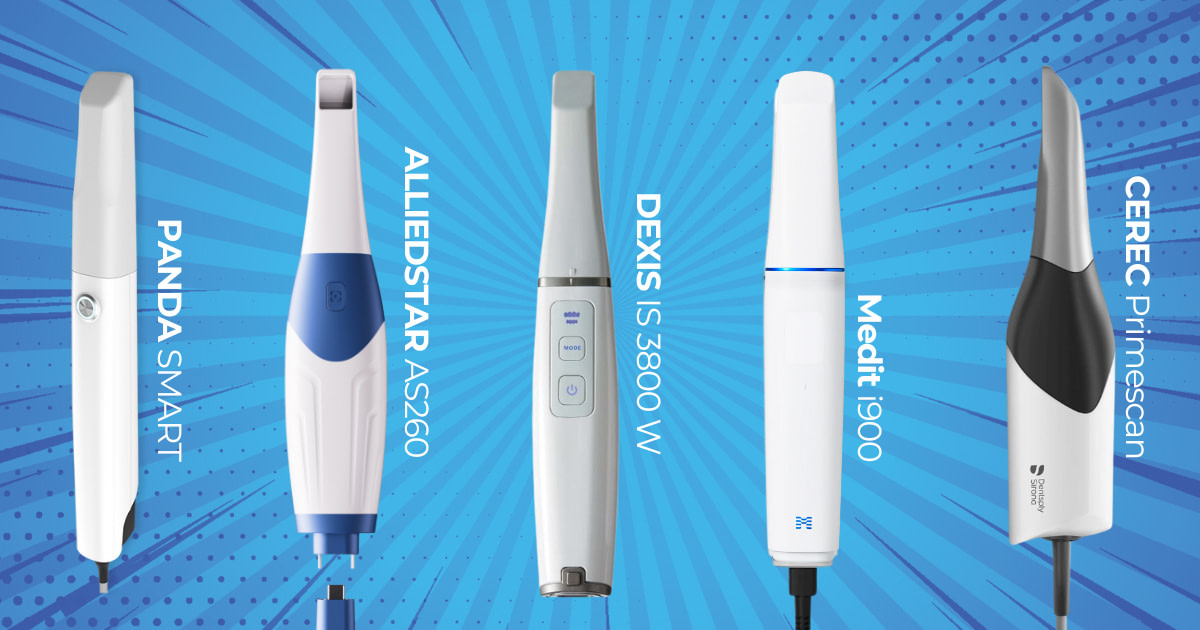
At the Institute of Digital Dentistry, we always aim to give our audience the content you want!
The team at iDD is over the moon to see how well-received these posts perform, so I’m happy to announce that our iDD Compares series will now be a weekly addition to our blogs!
Make sure you follow iDD across all our socials and subscribe to our newsletter to keep up to date with the latest Compares case we release, as well as other intraoral scanner (IOS) and 3D printer reviews and the latest announcements from the digital dentistry world.
I scanned this patient, the same 47 crown prep, with five different scanners on the same day:
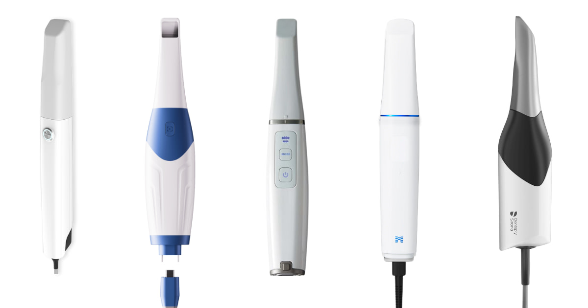
From left to right: Panda Smart, Alliedstar AS 260, DEXIS IS 3800 W, Medit i900, and CEREC Primescan.
Keep reading to see the results of the individual scans - pictures of the color scan, exported STLs, tessellated mesh, and a close-up of the prep margin.
Individual Scans in their Native Software
Every intraoral scanner has its own scanning software.
Almost all scanners can remove scan artifacts such as movable soft tissue, cheeks, and tongue through AI, with some performing better than others.
Interestingly, each scanner software captures color (called texture) differently. Below, you can see what the native scan images look like for each scanner.
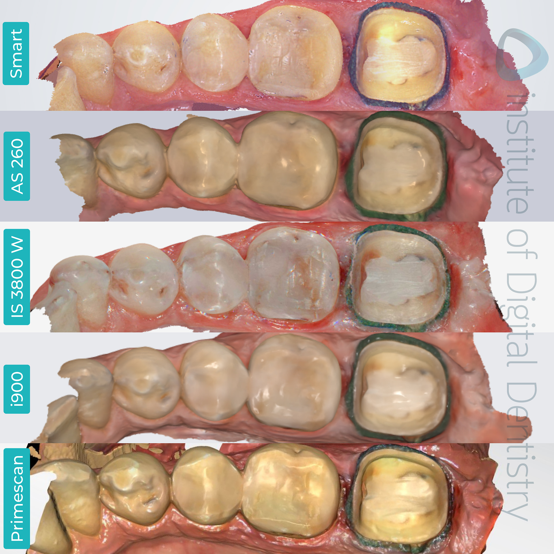
The processed, colored scans of the same tooth preparation were captured using five different scanners, as previewed in their native software.
At first glance, we can immediately identify that the AS 260 scan is the darkest out of the five IOS scans. This is closely followed by the i900 and IS 3800 W - although it’s important to note that DEXIS can toggle the brightness/light on and off within its native software.
The Panda Smart and the DEXIS IS 3800 W IOS capture more photorealistic scans and color texture, a staple that almost every Chinese IOS on the market possesses.
i900 has the classic Medit texture, with a high resolution and sharp margins. In contrast to the Chinese IOS, Medit’s scan is more of a cartoon-like aesthetic than the photo-realistic approach counterparts of Panda and DEXIS.
CEREC Primescan displays a more saturated, yellow hue than the other scans of the AS 260, i900, and IS 3800 W.
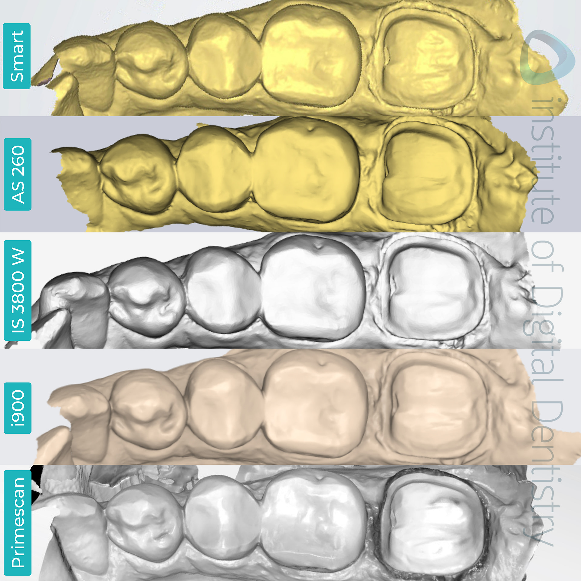
The processed monochromatic scans of the same tooth preparation were captured using five different scanners, as previewed in their native software.
Monochromatic scans can also be taken and previewed in their native software. By looking at scans in monochrome, one can get a better view of the prep's quality.
There aren’t usually too many major differences between the monochrome scans. However, Primescan does display more details on the prep quality in this view.
Exported Scans in Third-Party Software
All intraoral scanners have an open architecture that allows scans to be exported and sent to labs.
These scans are usually exported in three various formats - STL, PLY or OBJ files.
STL files are exported as monochromatic scans, whereas OBJ and PLY files store color (texture).
Not all intraoral scanners can export OBJ and PLY files, whereas STL files are widely used as the industry's default setting.
This particular set of scanners is capable of exporting scans in these formats:
- Medit i900 - STL, PLY and OBJ
- Panda Smart, Alliedstar AS 260, and DEXIS IS 3800 W - STL and PLY
- CEREC Primescan - STL only (unless connected to a lab via DS connect and then PLY can be exported lab side)
Intraoral Scanner | STL | PLY | OBJ |
|---|---|---|---|
Panda Smart | 12.3 MB | 10.7 MB | - |
Alliedstar AS260 | 4.2 MB | 1.7 MB | - |
DEXIS IS 3800 W | 4.4 MB | 2 MB | - |
Medit i900 | 4.9 MB | 2 MB | 4.9 MB |
CEREC Primescan | 15.2 MB | - | - |
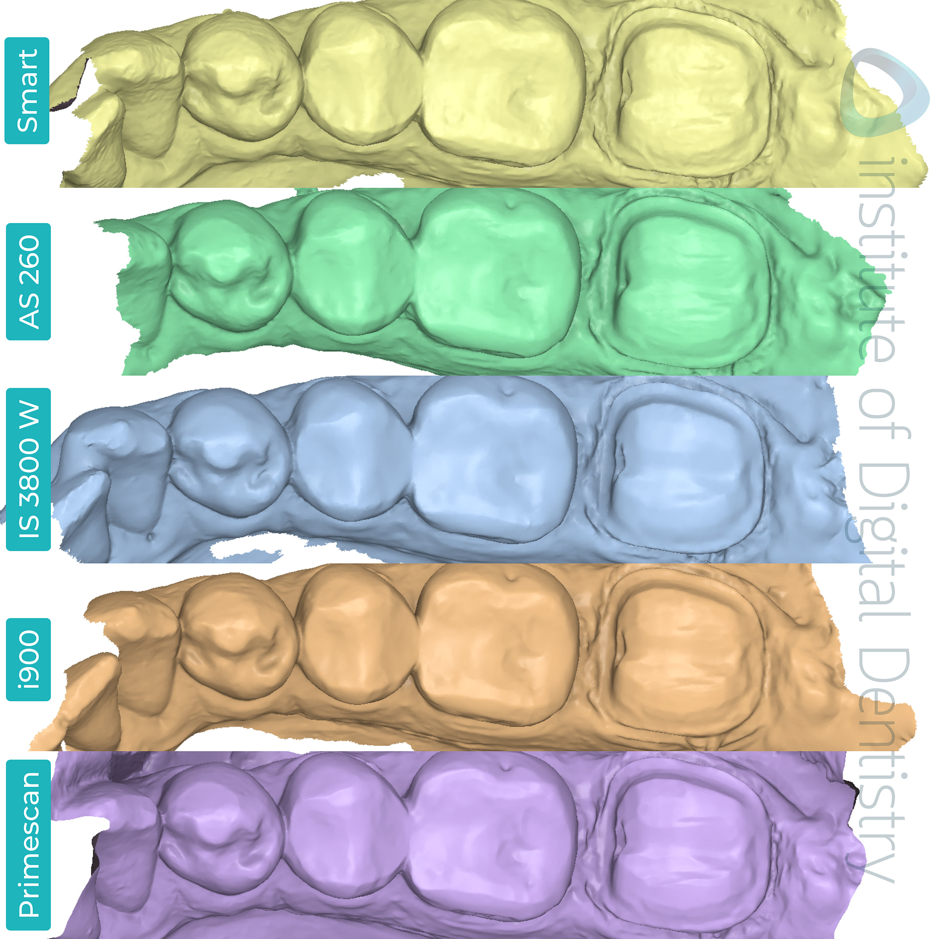
All scans were exported in an STL format and previewed in the Medit Design app.
When we look at the tessellated mesh of the scans from the various IOS devices, we can see that DEXIS’ IS 3800 W has the least dense tessellation mesh compared to Panda Smart’s and CEREC’s Primescan scans, which possess the densest tessellation mesh.

Studies investigating the clinical significance of mesh density have yet to be conducted from our understanding. It is important to note that a denser mesh does not necessarily indicate a ‘better scan.’
Dentsply Sirona’s CEREC Primescan is commonly seen as one of the gold standards of intraoral scanners (IOS) throughout the dental community, with it being regarded as one of the most accurate on the market.
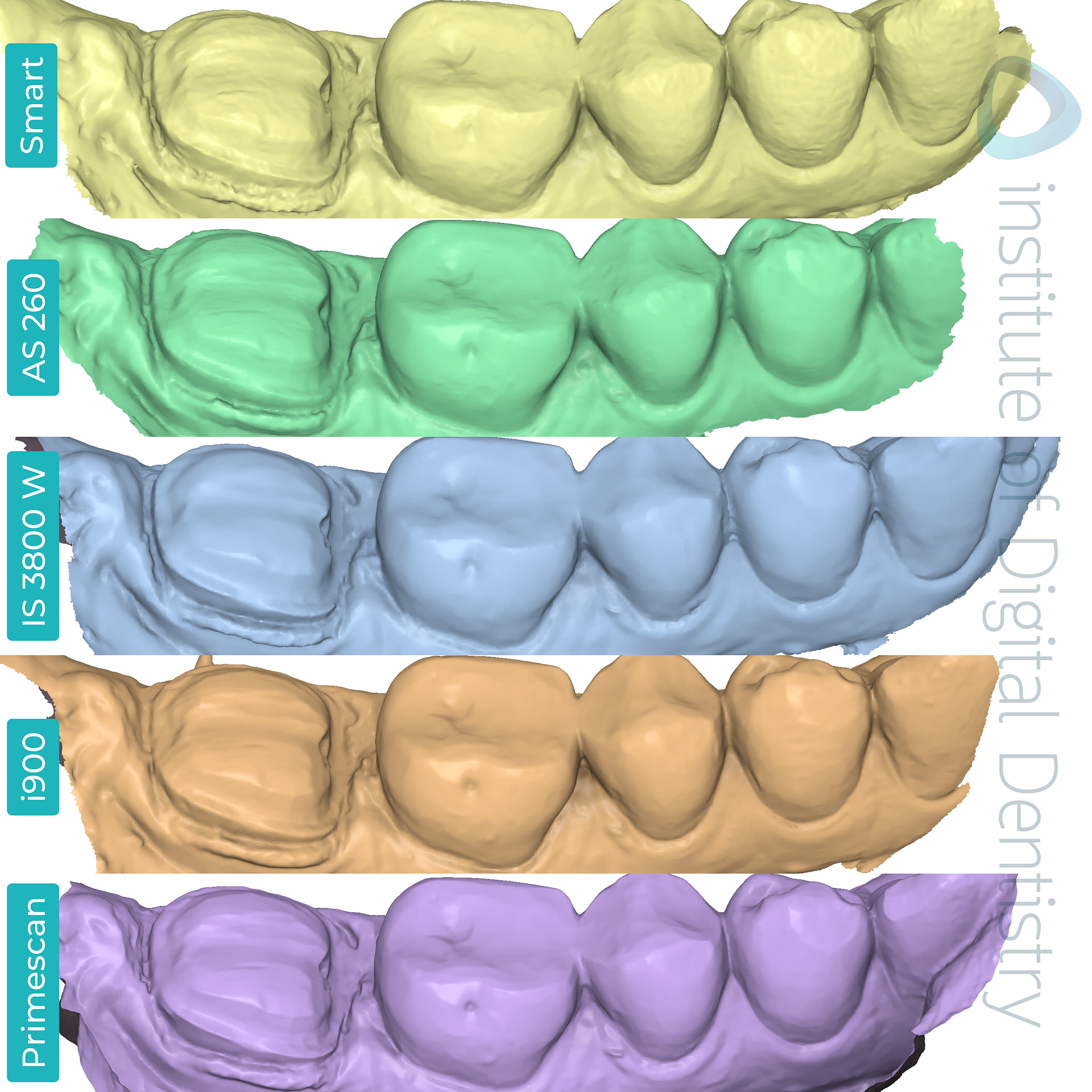

When viewing the preps buccally and lingually, we see that all scans captured the margin lines well. Primescan did capture it the smoothest with ever so slightly more definition.
To compare the deviations between the scans, we used CEREC Primescan as our point of reference. We aligned all five scans on top of each other to see if there were any major deviations that would be clinically significant.

Deviation map of the scans as compared to CEREC’s scan and sectional view. There seems to be minimal deviation around the prep area.
Based on the colored deviation key, we can see that the scan meshes are for the majority within 50 microns compared to the scan taken with CEREC Primescan.
However, we can see that there is some deviation between the scans as represented by the patches of yellow.

Deviation map of the individual scans compared to CEREC’s Primescan as our reference scan.
Conclusion
When it comes to capturing the details of restoration preparation, it's worth noting that the precision may vary somewhat based on the clinician's scanning technique and the specific scanner used.
This variation can affect the final restoration margin that the dental lab technicians create.
We're really interested to hear your take on this!
Leave a comment below and let us know which intraoral scanner you'd like to see us put head-to-head next.

