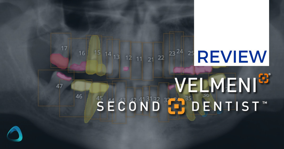
In recent years, artificial intelligence has been making significant contributions to various aspects of dentistry, with radiographic analysis being a prime area of focus. One of the emerging players in this field is Velmeni's Second Dentist platform, an AI-assisted tool designed to help with dental 2D and 3D radiograph interpretation.

If you've been following our reviews, you may remember our recent review of Pearl's Second Opinion AI platform. Both Pearl and Velmeni are at the forefront of bringing AI assistance to dental radiography, each with its unique approaches and features.
As the dental industry continues to evolve with technology, it's crucial to examine these innovations critically to understand their potential impact on clinical practice. Equally important is to remember that AI is a tool to assist, not replace, professional judgment. Dental professionals must be confident in their radiographic interpretation skills to effectively use and evaluate AI-assisted platforms.
In this review, we'll take a closer look at Velmeni's Second Dentist features, examining how they function and considering their potential utility in a dental practice setting. We'll explore the platform's interface, AI detection capabilities, image manipulation tools, and additional features for case presentation and documentation.
Our goal is to provide an unbiased view of what Second Dentist offers, helping dental professionals decide whether this tool might enhance their diagnostic processes and patient care.
It's worth noting that while AI in dentistry shows promise, its effectiveness can vary based on factors such as image quality, the specific conditions being detected, and how well the system integrates with existing workflows. With this in mind, let's dive into the features of Velmeni's Second Dentist platform and consider how it might fit into modern dental practice.
Having extensively tested Second Dentist in clinical practice, this review will provide you with an unfiltered, hands-on perspective of its strengths and limitations. As always, this is an independent review - Velmeni had no involvement in its creation or content. My goal is to help you make an informed decision about whether this platform could benefit your practice and patients.
Enjoy the review.
Download The Full Review
Download the full page PDF to get a full copy of this article to read later.
Get a high resolution printable copy of this review.
What is Second Dentist?
Velmeni is the company, and the platform is called Second Dentist. It is a browser-based platform that integrates with your imaging software or PMS to automatically upload any 2D or 3D radiographs taken.
While the fundamental concept might seem familiar - AI-assisted radiograph interpretation - Second Dentist brings some distinctive features to the table that immediately set it apart from its competitors.
The most notable distinction is Second Dentist's dual capability in analyzing both 2D and 3D radiographs. This is relatively unique in the current market where most platforms focus exclusively on 2D analysis. Currently, the only other platform that can analyse both 2D and 3D x-rays is Diagnocat.
Another nice feature of Second Dentist is its flexibility in image input. Unlike other companies (like Pearl) that upload and analyze x-rays via software integration only, Second Dentist offers both automated and manual upload options. In other words, you can either have radiographs automatically populated through your imaging software integration or manually upload any radiographs directly through the web platform. This flexibility proves particularly valuable when dealing with external referrals or when reviewing historical records from other practices.
Screenshot
At its core, the platform analyzes radiographs to identify and highlight potential areas of concern, covering all the standard elements we've come to expect from dental AI diagnostics such as caries detection, calculus presence, bone loss measurements, and periapical radiolucencies. However, it extends beyond these basics with its 3D capabilities, offering pathology detection and anatomical structure identification in CBCT scans.
The comapny says the platform was refined through direct input from over 50 practicing dentists, clinical validation from Velmeni's dental board members, and beta testing with 1,300 dentists in real clinical settings. This extensive field testing has yielded impressive validation statistics - over 90% of participating dentists reported that the AI technology would be beneficial in their practice and expressed willingness to adopt it.
Like other players in the dental AI space, Velmeni positions Second Dentist as an assistive tool rather than a replacement for professional judgment. The name itself - "Second Dentist" - cleverly emphasizes this supportive role while subtly differentiating itself from competitors' "Second Opinion" messaging.
The crucial question, of course, is how well these features perform in real-world clinical settings - something we'll explore in detail throughout this review. As with any AI tool in healthcare, it's essential to approach Second Dentist as an aid to, rather than a replacement for, professional judgment. Let's dive deeper into how this platform performs in daily practice.
FDA Clearance
In a milestone for dental AI technology, Velmeni secured FDA 510(k) clearance on September 3, 2024, for their VELMENI for DENTISTS (V4D) platform. This breakthrough positions Velmeni as one of the first non-US based dental AI companies to receive FDA approval for pathology detection in panoramic X-rays, alongside bitewing and periapical radiographs. The significance of this approval is noteworthy, as it places Velmeni in an elite group of dental AI companies with regulatory approval, joining established players like Overjet and Pearl.
The scope of the FDA clearance is comprehensive, covering detection of dental caries, identification of fillings and restorations, recognition of fixed prostheses, implant detection, and analysis across multiple radiograph types including bitewing, periapical, and panoramic radiographs. The clearance specifically applies to permanent teeth in patients 15 years and older. This regulatory milestone builds upon Velmeni's existing international certifications, including Canadian MDEL (Medical Device Establishment License) and New Zealand MEDSAFE approvals.
Velmeni Interface and Image Management
Second Dentist's interface tries to strike a balance between functionality and simplicity, though it takes a notably different approach from Pearl's sleek, minimalist design.
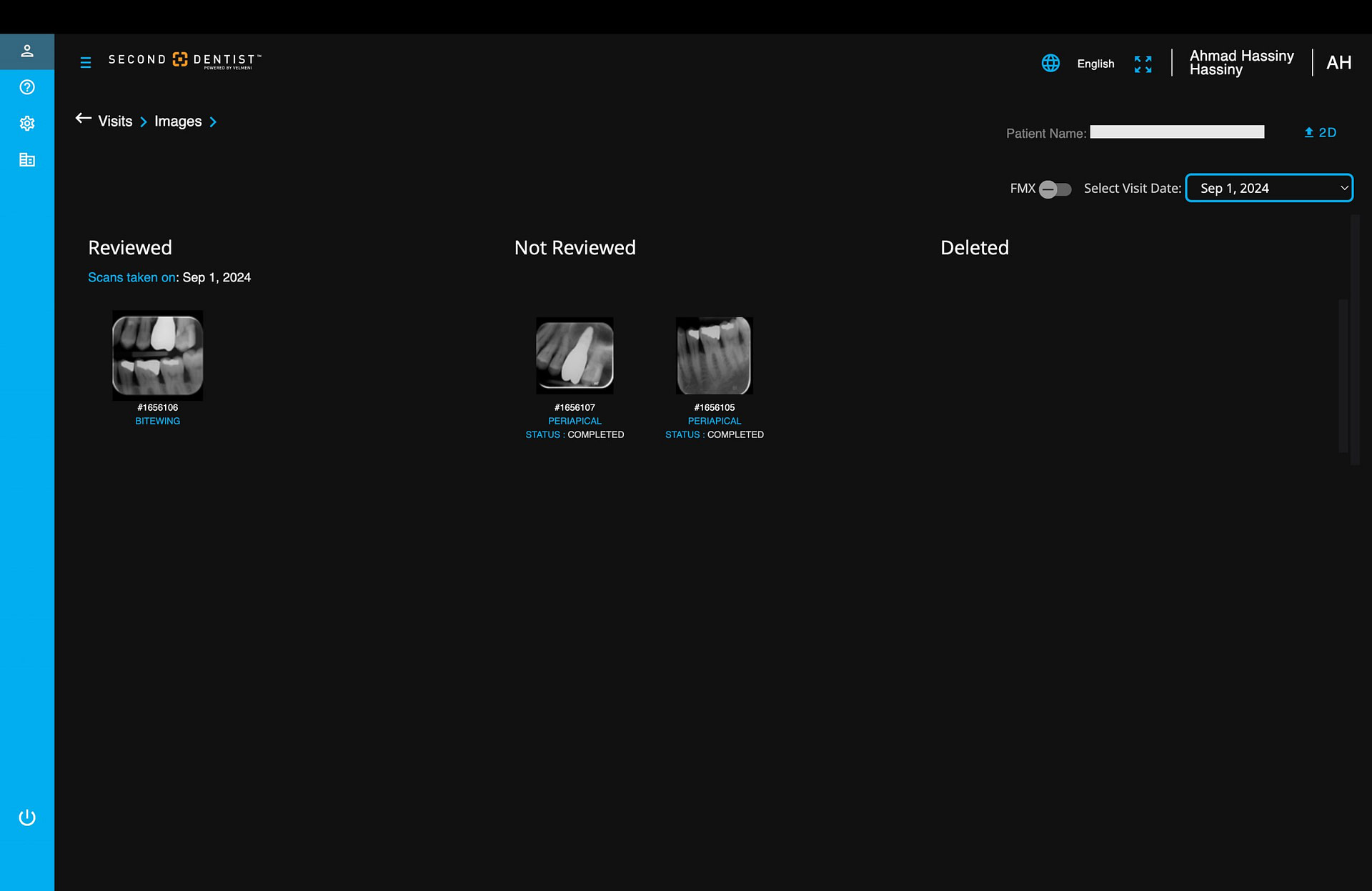
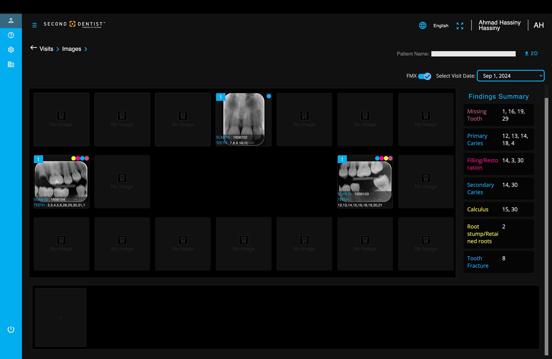
Upon logging in through your web browser, you're presented with a dashboard that organizes patient files chronologically, with the most recent scans displayed first. Clicking on any of the 'visits' buttons will show you all the radiographs populated into the software for that patient on that patient visit.

The interface uses a tile-based layout for radiographs where each patient's radiographs are grouped together, making it simple to access recent cases quickly. Once, again Second Dentist allows you to manually upload radiographs directly through the browser interface. The drag-and-drop upload functionality is straightforward, accepting both 2D and 3D DICOM files without fuss. The platform automatically attempts to arrange serial radiographs in chronological order, making it easier to track changes over time.
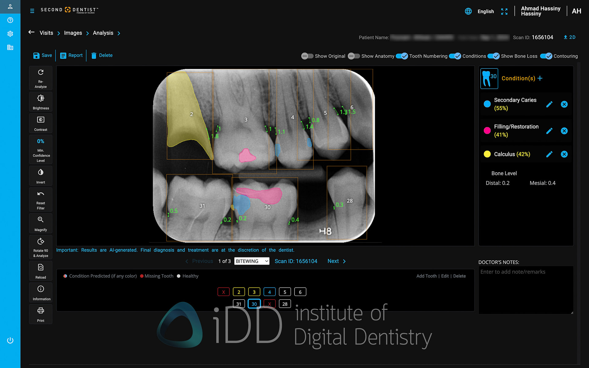
Navigation within the platform feels mostly intuitive, if somewhat utilitarian. The image viewer occupies most of the screen real estate, with AI detection controls arranged in a toolbar along the top with simple on and off toggles. A side panel includes standard image manipulation tools - brightness, contrast, and zoom controls etc.
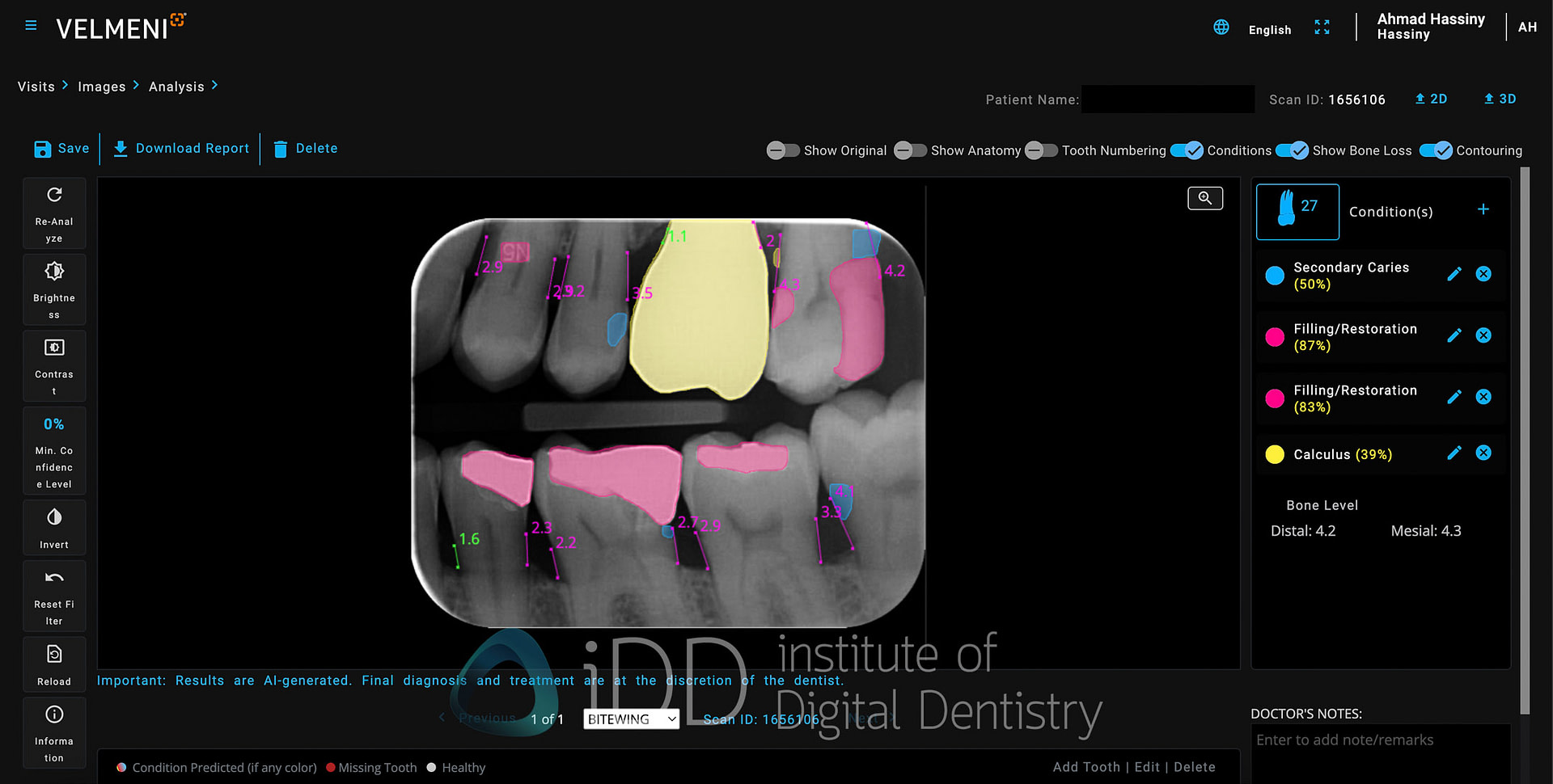
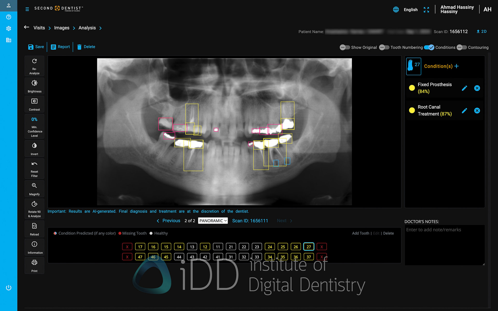
The software automatically segments each x-ray into tooth numbers. Each type of detection (caries, bone loss, etc.) can be toggled individually by hovering over it, preventing the screen from becoming cluttered with markings. There is also a colour-coded summary at the bottom that indicates what detection has been found for each tooth.
Screenshot
When you click on any tooth, the window on the right provides additional details about the finding, including measurement data and confidence levels. The ability to edit or dismiss AI findings is straightforward, acknowledging that professional judgment should always take precedence over automated detections.
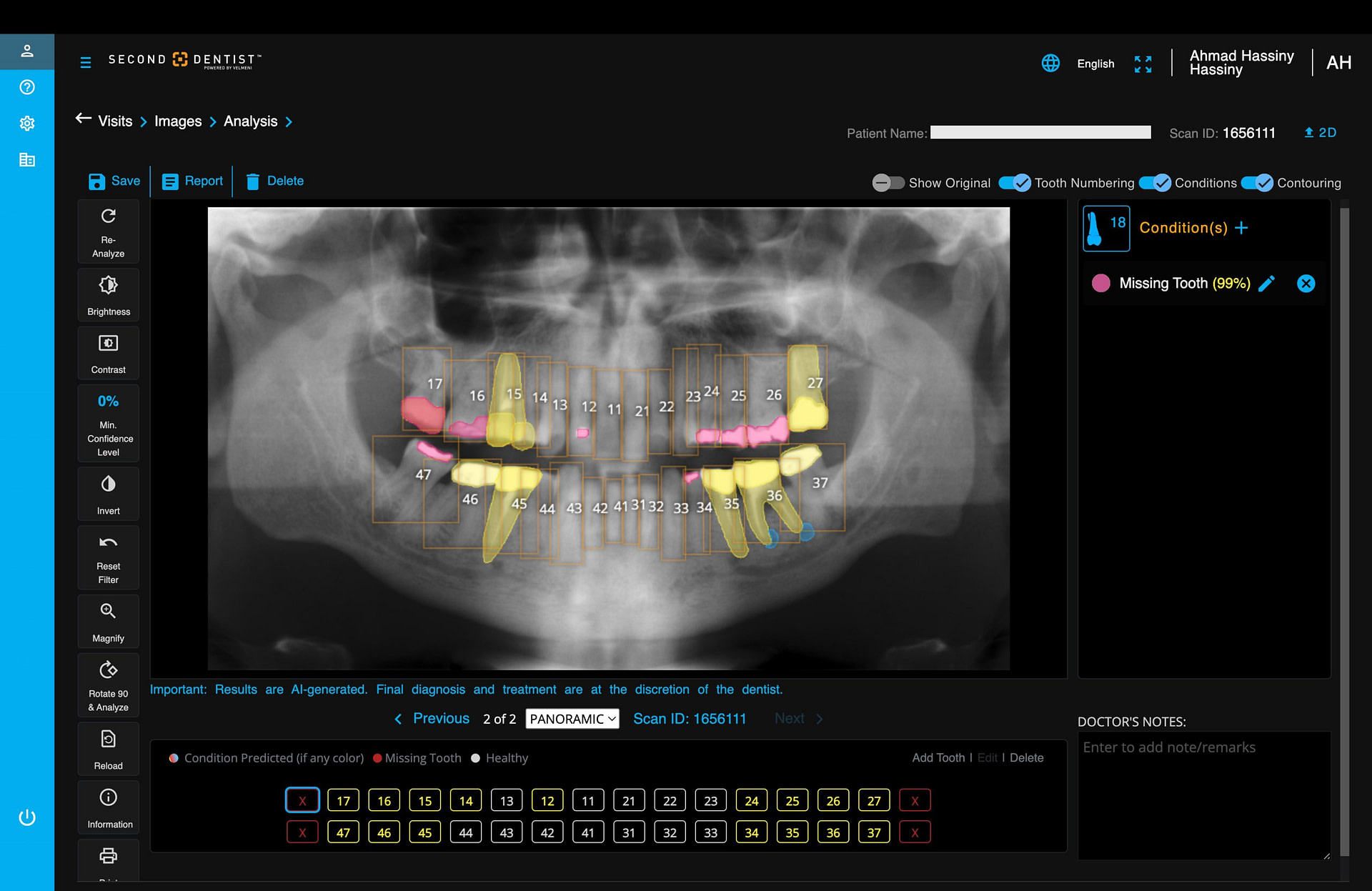
One of Second Dentist's useful features is its ability to toggle between different visualization styles for AI findings. The platform offers two display modes: contour shading and box indicators. The contour shading option, which I find far more effective, fills in detected areas with semi-transparent colors that follow the natural contours of the findings. This creates a cleaner, more intuitive visualization that makes it easier to understand the extent and shape of detected pathologies.
The alternative box indicator mode simply places colored squares around areas of interest. While this might appeal to some users, I find it can make the radiograph appear cluttered, especially when multiple findings are present in close proximity. The boxes can sometimes obscure subtle details in the surrounding areas.
You can easily switch between these visualization styles through a simple toggle in the interface. This flexibility allows users to choose the display method that best suits their preferences, though in my experience, the contour shading provides a more refined and less distracting way to visualize AI detections.
Although there are some minor quirks, such as the interface occasionally feels overloaded with options and the UI does feel a bit more dated than Pearl, overall Second Dentist's interface successfully delivers on its core promise: providing a functional platform for AI-assisted radiograph analysis that can handle both 2D and 3D imaging. While it may not win any awards for revolutionary design, it gets the job done efficiently once you're familiar with its workflow.
AI Detections
The cornerstone of Second Dentist's offering is its AI-powered detection system for both 2D and 3D radiographs. Second Dentist aims to assist dental professionals in identifying a range of dental conditions by automatically highlighting areas of potential concern.
For 2D radiographs, the system focuses on five key areas of detection, each utilizing distinct visual markers and color coding for clear identification. These six areas being:
- Caries
- Bone levels
- Calculus
- Restorations
- Periapical Radiolucencies
Let's break down some of these key features:
Caries Detection
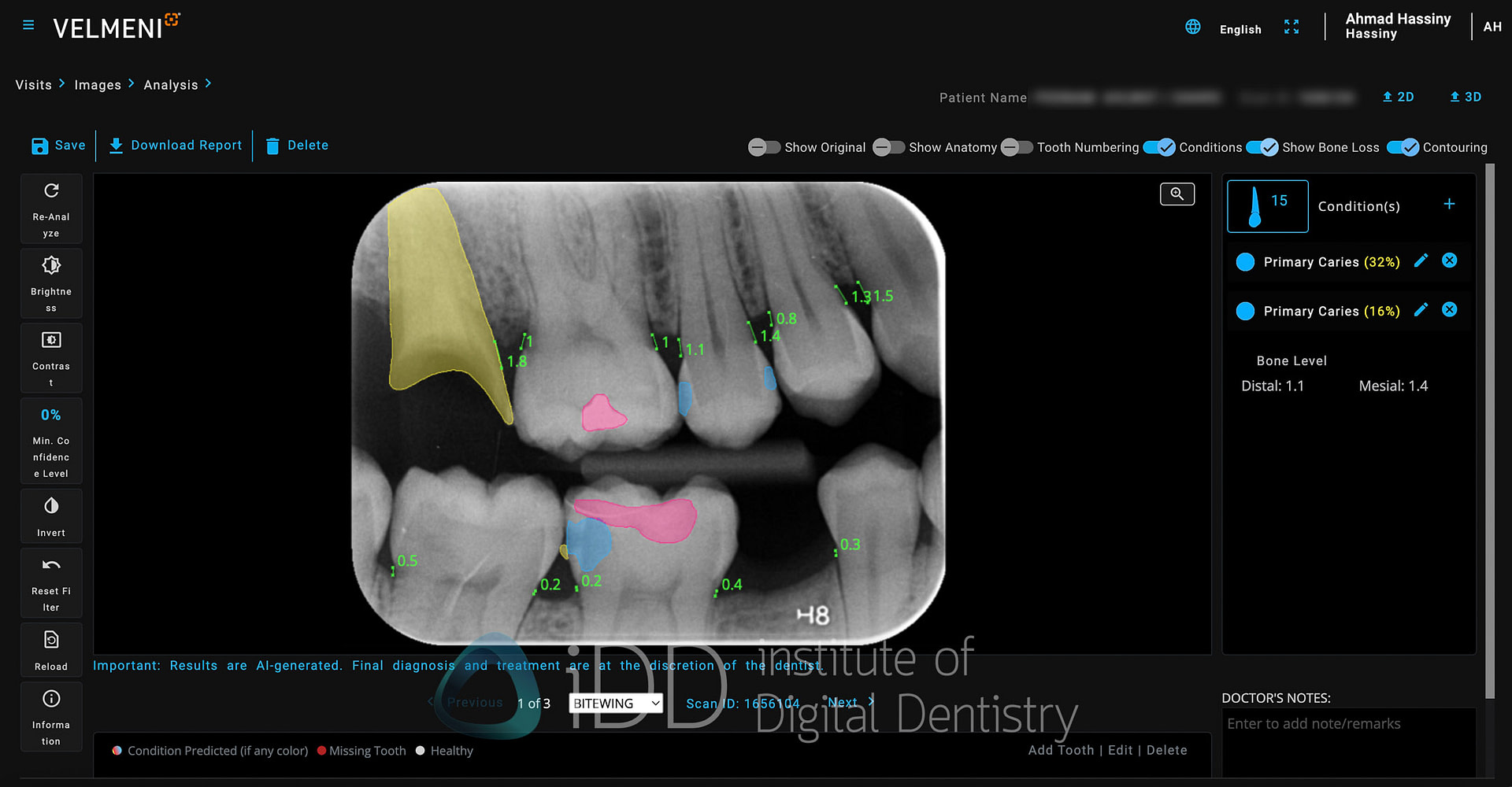
Second Dentist takes a straightforward approach to caries detection with a simple marking system. The platform uses a single color - blue - to highlight areas of suspected decay, whether it's primary caries in untreated teeth or secondary caries around existing restorations. The blue markers clearly stand out against the grayscale of the radiograph, drawing attention to suspicious areas that warrant closer examination.
In daily practice, I've found this visualization particularly effective for patient communication. The blue highlights make it easy to explain findings to patients. Whether pointing out early decay between teeth or showing secondary caries beneath an existing restoration, the consistent blue marking system helps maintain clear communication.
However, Velmeni has thoughtfully designed the system to complement rather than replace clinical judgment. While the AI excels at highlighting suspicious areas that might be easily missed during initial examination, the platform emphasizes that these findings should be considered alongside clinical examination findings. Each AI detection can be evaluated and either accepted or rejected by the dentist, maintaining the crucial role of professional expertise in diagnosis and treatment planning.
While other AI platforms use multiple colors or the severity of the decay, Second Dentist's single-color approach doesn't seem to have this feature yet.
Bone Measurements
Second Dentist's approach to bone level assessment uses a straightforward two-color system paired with automated measurements to streamline the detection of bone loss.
The AI calculates the distance from the alveolar bone crest to the cementoenamel junction (CEJ), providing standardized measurements that help clinicians quickly identify areas of concern.
The platform keeps things simple with a two color-coding system: green lines indicate healthy bone levels (up to 2.5mm from CEJ), while pink lines signal potential bone loss (anything exceeding 2.0mm). This clear visual distinction makes it easy to spot areas requiring attention at a glance.
Once again, Second Dentist maintains transparency about the limitations of radiographic bone measurements. The system clearly acknowledges that image angulation can affect measurement accuracy - a reminder that these findings should be correlated with clinical probing depths and other diagnostic indicators. This reinforces the platform's intended role as a diagnostic aid rather than a standalone diagnostic tool.
The real strength of this feature lies in its simplicity and efficiency. By automating the initial measurements with a clear binary indication of health versus disease, while still allowing for professional oversight, it streamlines the periodontal assessment process without sacrificing clinical accuracy. There is no colour indication for severity however.
Calculus Detection
Second Dentist uses yellow indicators to highlight areas where radiographic calculus is detected. The AI analyzes radiographs for the characteristic appearance of calculus - those radiopaque deposits along root surfaces - and marks them for easy identification.
This automated detection is particularly useful for identifying subgingival calculus that might not be immediately visible during clinical examination. The yellow markers provide a quick reference during treatment planning, helping identify areas that will require scaling during hygiene appointments.
The feature serves both clinical and educational purposes. For hygienists, it offers a useful overview of calculus distribution before beginning treatment. For patient education, the clear visual indicators help demonstrate the presence of calculus deposits and explain the need for professional cleaning.
As with all AI detections in Second Dentist, these findings are meant to supplement rather than replace clinical examination and professional judgment. The system allows practitioners to verify, modify, or dismiss any detected areas based on their clinical assessment.
Restorations
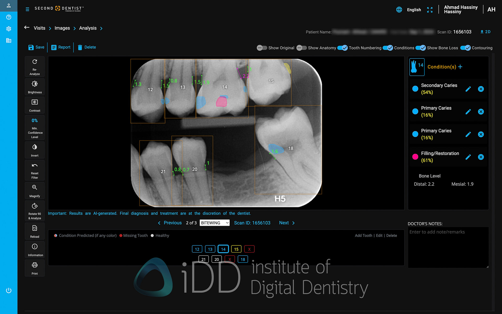
Second Dentist uses a two-color system for marking existing dental restorations on radiographs. Fixed prosthetics like crowns and bridges are highlighted in yellow, while direct restorations such as composite or amalgam fillings are marked in pink.
This clear visual distinction between restoration types helps quickly identify different types of existing dental work during radiograph interpretation. The color-coding also proves useful during patient examinations, making it easy to point out different types of restorations when discussing treatment history or planning future work.
The AI detection includes both complete restorations and partial coverage restorations, marking any radiopaque dental materials present in the radiograph. As with other detections, these markings can be toggled on or off as needed during examination, and practitioners can modify or remove any incorrectly marked areas.

Download The Full Review

Download the full page PDF to get a full copy of this article to read later.
Get a high resolution printable copy of this review.
Periapical Radiolucency Detection
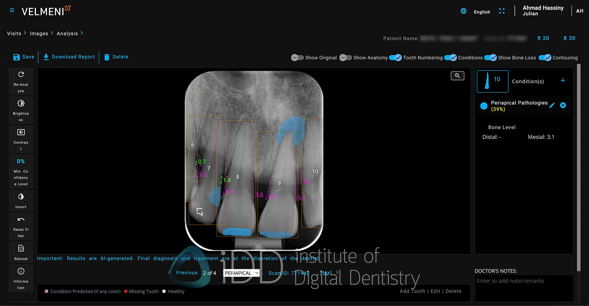
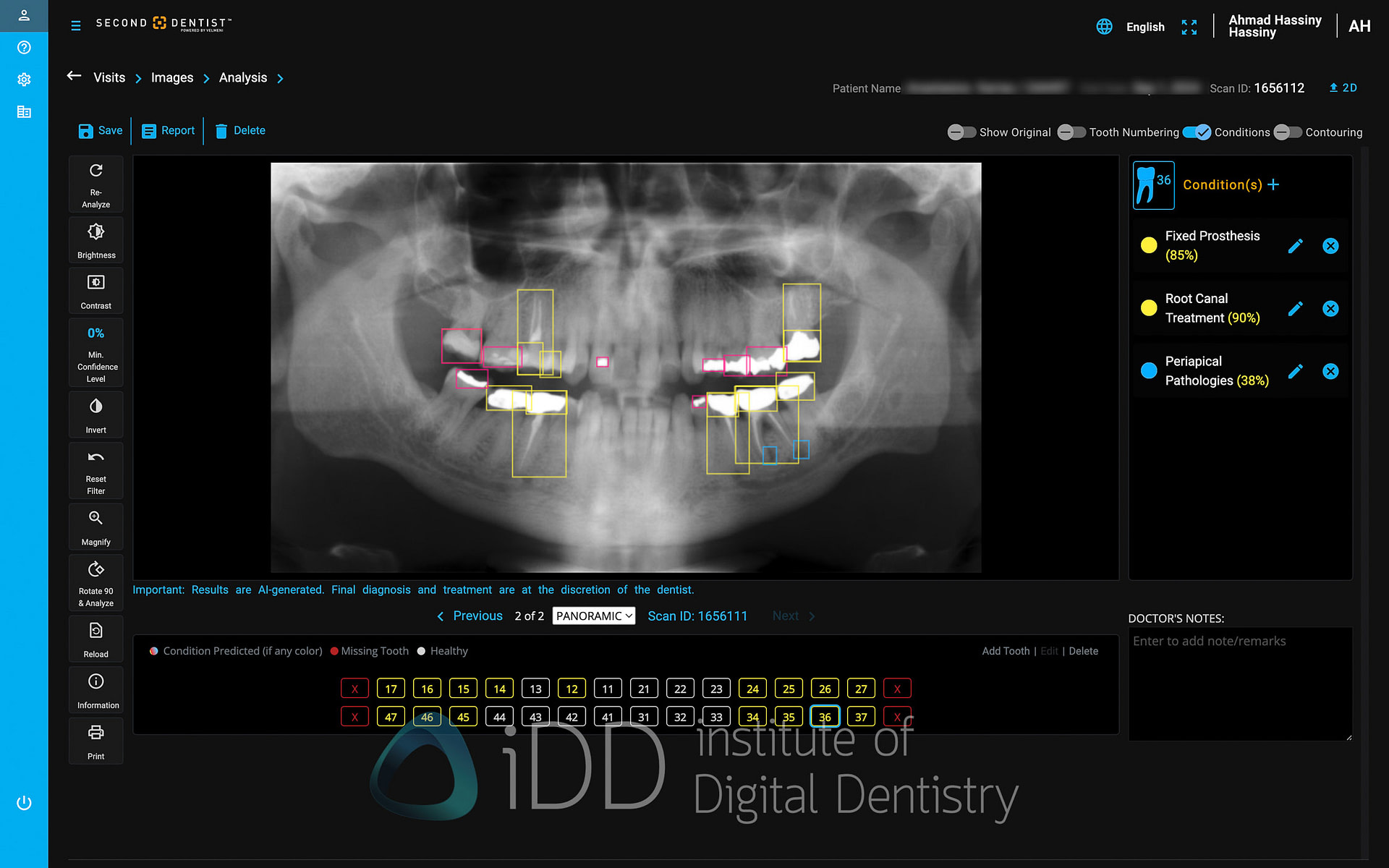
Second Dentist marks periapical radiolucencies with blue indicators, highlighting areas of potential pathology around root apices. The AI analyzes the radiographic density around root tips to identify radiolucent areas that might indicate periapical inflammation or infection.
The blue markings help draw attention to these significant findings during radiograph interpretation. This automated detection can be particularly useful for identifying subtle periapical changes that might be easily overlooked during initial examination, especially in multi-rooted teeth or anatomically complex areas.
As with all AI detections in Second Dentist, these findings serve as a screening tool rather than a definitive diagnosis. Each detection can be evaluated and modified by the practitioner based on clinical findings and professional judgment. The system allows practitioners to easily toggle these markings on or off, and to add or remove detections as needed during examination.
Tooth Segmentation
Second Dentist incorporates robust tooth segmentation technology that automatically identifies and outlines individual teeth in both 2D and 3D radiographs. The AI precisely delineates tooth boundaries, creating clear segmentation lines that separate each tooth from its neighbors.
This segmentation occurs automatically when radiographs are uploaded, requiring no manual input. The system accurately identifies tooth morphology, distinguishing between different tooth types and accounting for variations in positioning and overlap. Each segmented tooth is automatically numbered according to the universal numbering system, making it easy to reference specific teeth during examination and treatment planning.
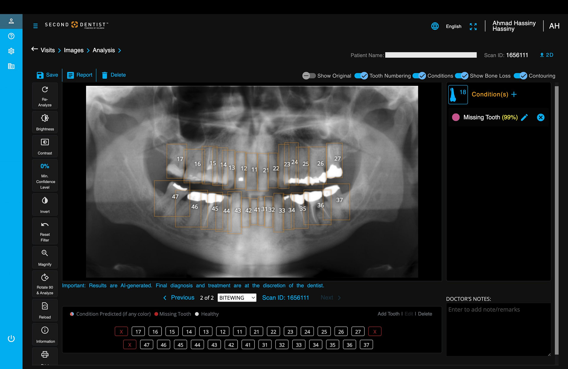
The segmentation proves particularly valuable when combined with other AI detections, as it helps localize findings to specific teeth and surfaces with greater precision. When viewing detected pathologies like caries or periapical lesions, the clear tooth boundaries provide important contextual reference points.
The accuracy of the segmentation is notably consistent, even in challenging cases with overlapped teeth or unusual anatomical variations. This reliable tooth separation helps maintain clarity in radiographic interpretation and improves the specificity of AI findings.
Sometimes AI Doesn't Get It Right
AI is fundamentally a tool, and those who expect perfect accuracy every time are approaching it incorrectly. I view Second Dentist primarily as a way to standardize diagnosis and enhance patient communication, not as an infallible diagnostic oracle.
During my time using Second Dentist, I've noticed a few instances where the AI either missed findings or made incorrect detections:

However, these limitations are expected and understandable. The key advantage of AI is its consistency - it doesn't experience fatigue, distraction, or variation in performance like humans do. It analyzes every radiograph using the same objective criteria each time.
Would I rely entirely on Second Dentist's AI detections? Absolutely not. But that's not its intended purpose. It serves as a valuable screening tool and communication aid while acknowledging that final diagnostic decisions require professional judgment and consideration of multiple clinical factors beyond radiographic appearance alone.
One of the most intriguing aspects of dental AI that I've discovered is how different AI platforms can analyze the same radiograph and produce notably different results. Running identical radiographs through Pearl, Velmeni, and other AI systems often yields varying detections and interpretations.
For example, in assessing bone levels, the measurements can vary between platforms. What one system confidently marks as calculus, another might overlook completely. Even in seemingly straightforward tasks like identifying restorations or carious lesions, there can be surprising discrepancies between platforms.
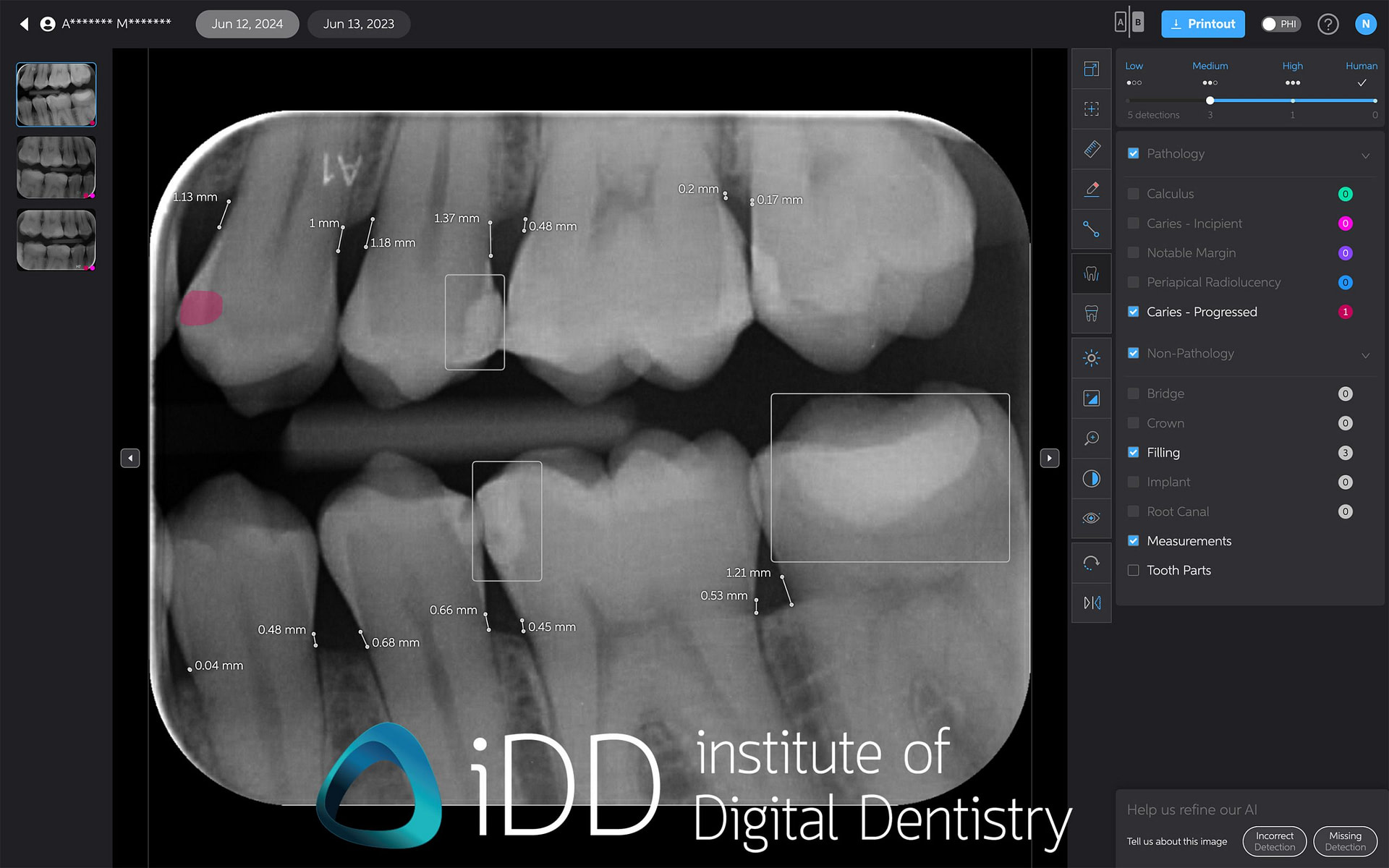
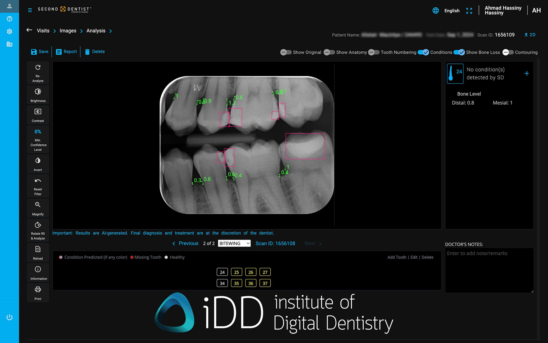
This variation highlights the current state of AI in dentistry. Just as different dentists might have varying opinions on borderline cases, these AI systems have been trained on different datasets and using different methodologies, leading to varying thresholds for what constitutes a positive finding.
This observation reinforces two important points: first, that AI should be viewed as a supportive tool rather than a definitive diagnostic oracle, and second, that professional judgment remains crucial in interpreting both the radiographs and the AI's suggestions. It's a fascinating reminder that we're still in the early stages of AI in dentistry, where even the definition of what constitutes a "correct" detection is still evolving along with the technology itself.
Case Presentation and Documentation
Second Dentist includes mulitple tools for case presentation and documentation. The presentation mode provides a clean interface for patient education, where AI findings can be toggled on or off to build explanations progressively. The tooth segmentation makes it particularly easy for patients to understand which teeth are affected.
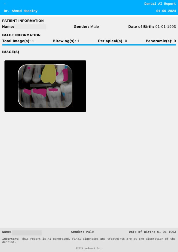
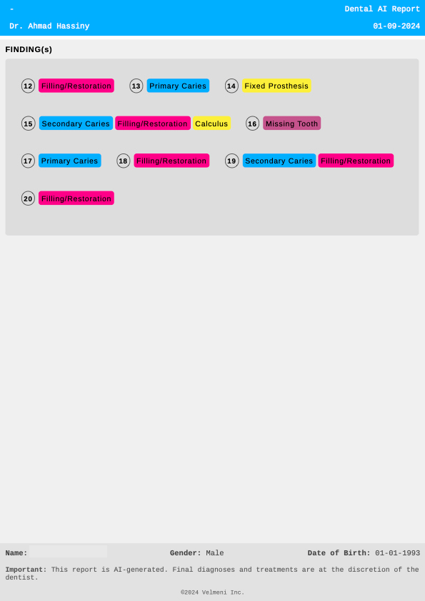
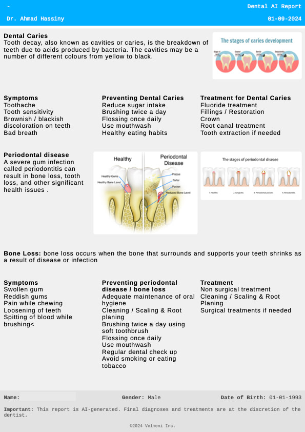
For documentation, the system generates reports of AI findings that can be exported as PDFs for patient records or referrals. All detections are automatically timestamped and logged, creating a searchable history of findings. The platform allows practitioners to add notes and annotations, which become part of the permanent record.
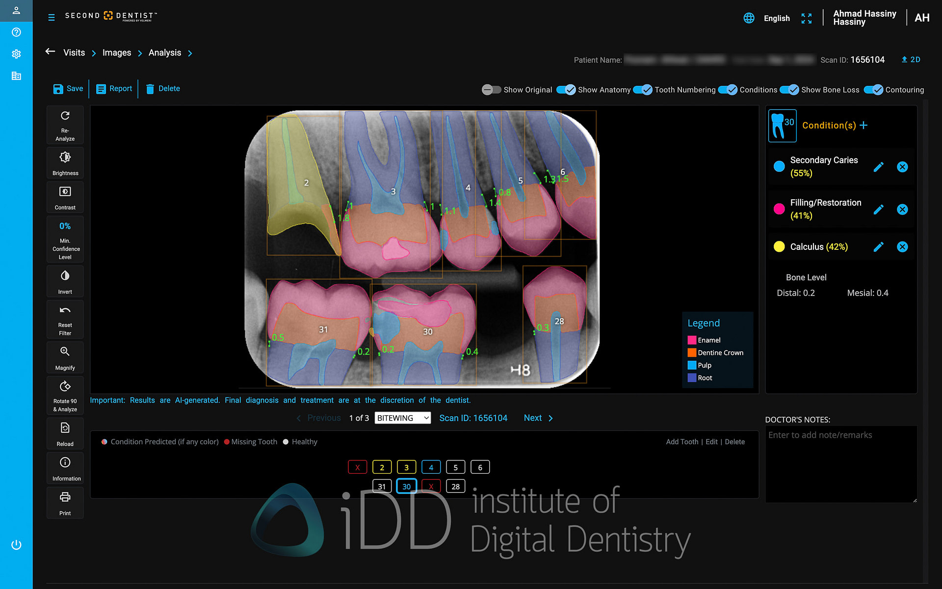
Distinguishing different tooth structures
Visually distinguishing different tooth structures helps patients better understand their conditions and proposed treatments.
Compare Mode
This feature allows side-by-side viewing of current and historical images. It is useful for demonstrating disease progression or treatment outcomes to patients. Each image in compare mode has its own toolbar, allowing for individual manipulation.
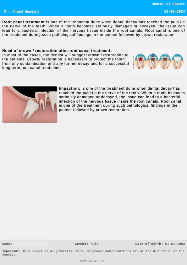
Printouts
The platform offers customizable printout options, which are helpful for patient handouts, referrals, or insurance claims. Users can input their practice's contact information, which is included in printouts. Anything discussed on Second Dentist can be printed and given to the patient.
Download The Full Review

Download the full page PDF to get a full copy of this article to read later.
Get a high resolution printable copy of this review.
Other features of Second Dentist
One of the strengths of Second Dentist's AI detection system is its flexibility. You can toggle different types of detections on and off, allowing for customized views depending on the focus of examination or patient education needs.
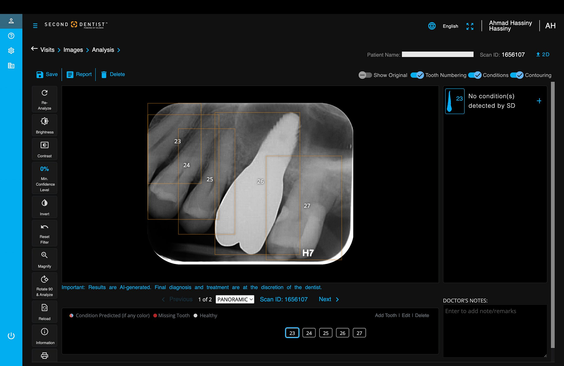
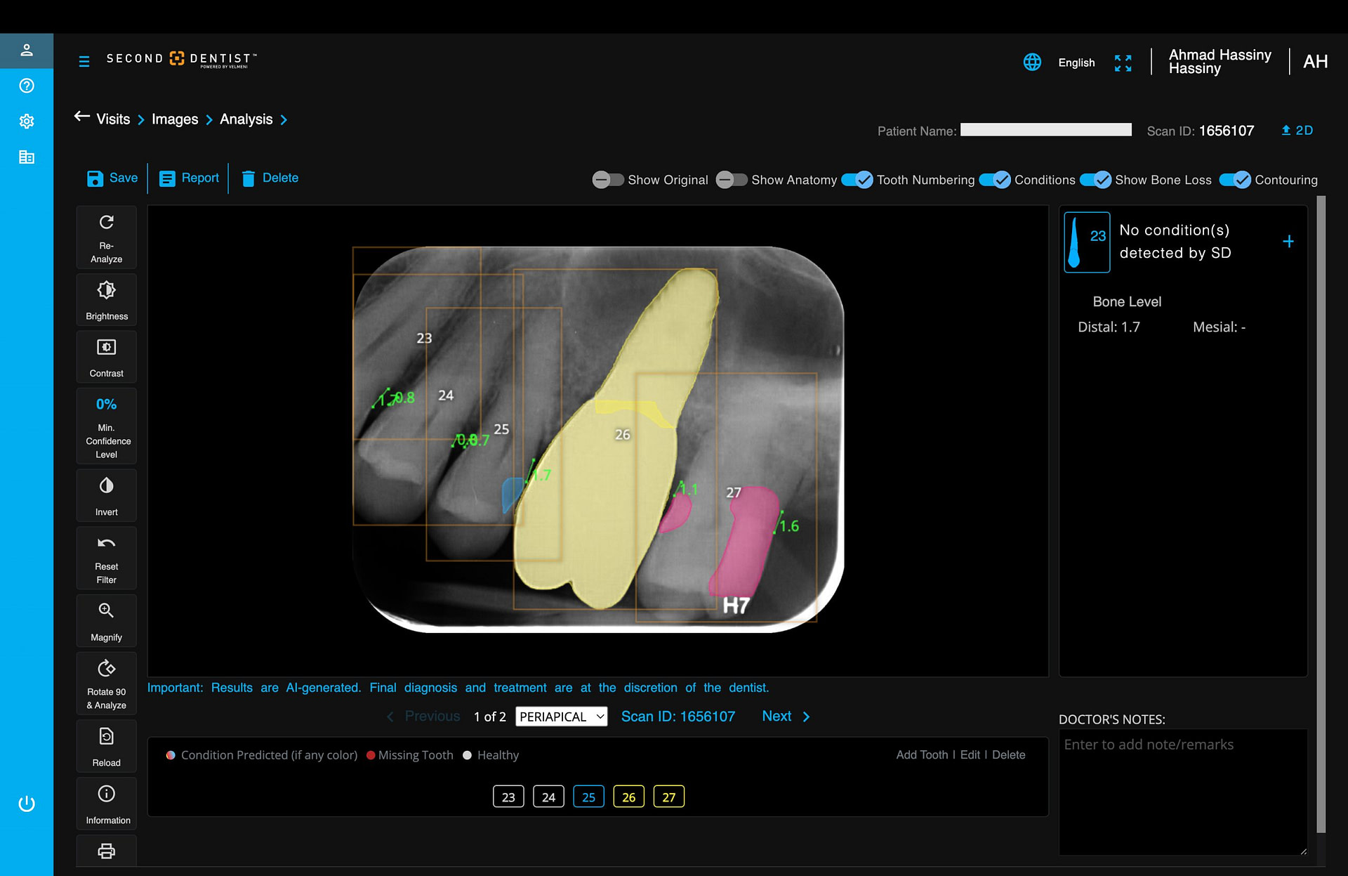
The system also provides an editing tool, allowing users to remove false positives or add detections the AI may have missed. This acknowledgment that AI is not infallible and that the dentist's judgment is paramount is a positive aspect of the entire system.
The Cost of Second Dentist
Second Dentist operates on a straightforward subscription model, priced at $399 USD per month. This subscription includes both the 2D and 3D radiograph analysis capabilities, making it a comprehensive solution for practices using various types of dental imaging.
The pricing positions Second Dentist competitively in the dental AI market. While this represents an additional monthly overhead for practices, the value proposition lies in its potential to enhance diagnostic consistency, improve patient communication, and streamline workflow efficiency.
Conclusion
Second Dentist represents an intriguing addition to the dental AI landscape, distinguishing itself through a unique combination of accessibility and capability. Its standout feature - the ability to analyze both 2D and 3D radiographs in a single platform - sets it apart in a market where most competitors focus solely on 2D analysis.
At $399 monthly, Second Dentist positions itself as a more cost-effective alternative to premium platforms that often exceed $500-600 per month. While its interface might appear more utilitarian compared to some higher-priced competitors, this simplicity contributes to its accessibility somewhat.
The platform offers some practical advantages that even premium competitors lack, most notably the ability to manually upload radiographs - a seemingly simple feature that proves invaluable when reviewing external cases or historical records. This flexibility, combined with its 3D analysis capabilities, helps offset its more basic interface.
As with any AI diagnostic tool, Second Dentist has its limitations. It can miss findings or make incorrect detections, serving as a reminder that these platforms should augment rather than replace professional judgment. However, for practices seeking to integrate AI into their diagnostic workflow without premium pricing, Second Dentist offers a compelling balance of features and affordability.
In essence, Second Dentist demonstrates that effective AI solutions don't necessarily require premium pricing. By focusing on core functionality while offering comprehensive 2D and 3D analysis capabilities, it presents a practical entry point for practices looking to integrate AI into their diagnostic workflow. Overall it is a noteworthy contender in the evolving dental AI landscape.
Ready to enhance your radiographic interpretation skills?
Our 2D Radiography Interpretation Course provides comprehensive training to boost your confidence and expertise.
Enroll now and take your diagnostic skills to the next level!

