iDD is back yet again with another intraoral scanner (IOS) comparison.
This time, Dr. Ahmad Al-Hassiny used two IOS to assess their performance when capturing images of the same implant patient, focusing on the scan body and the surrounding soft tissue.
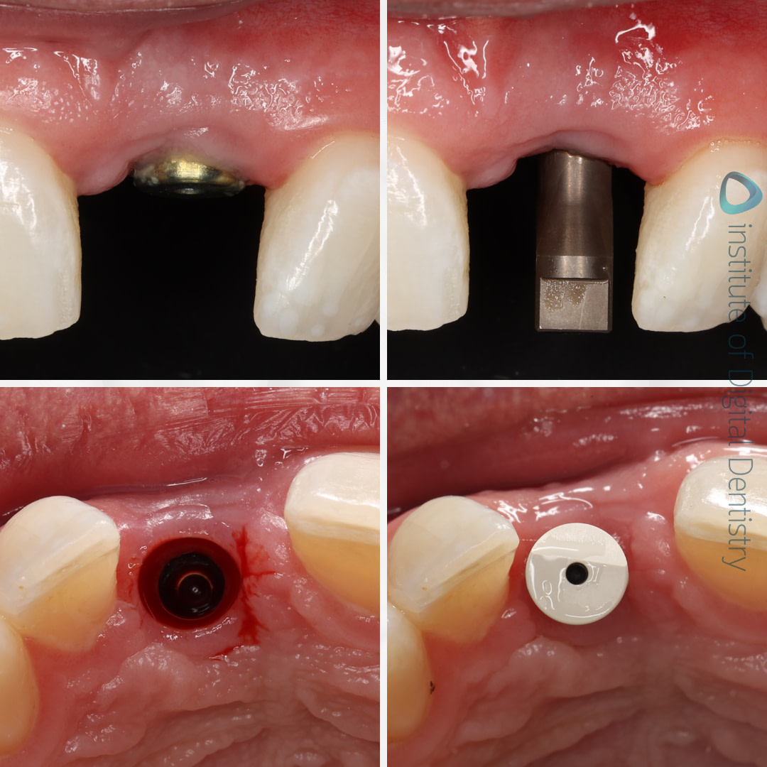
Photos of implant scan body.
Here at the Institute of Digital Dentistry, we are fortunate to have access to a wide selection of IOS devices. For this particular case comparison, scans were taken using Medit’s i700 Wireless and Runyes’ 3DS 3.0.
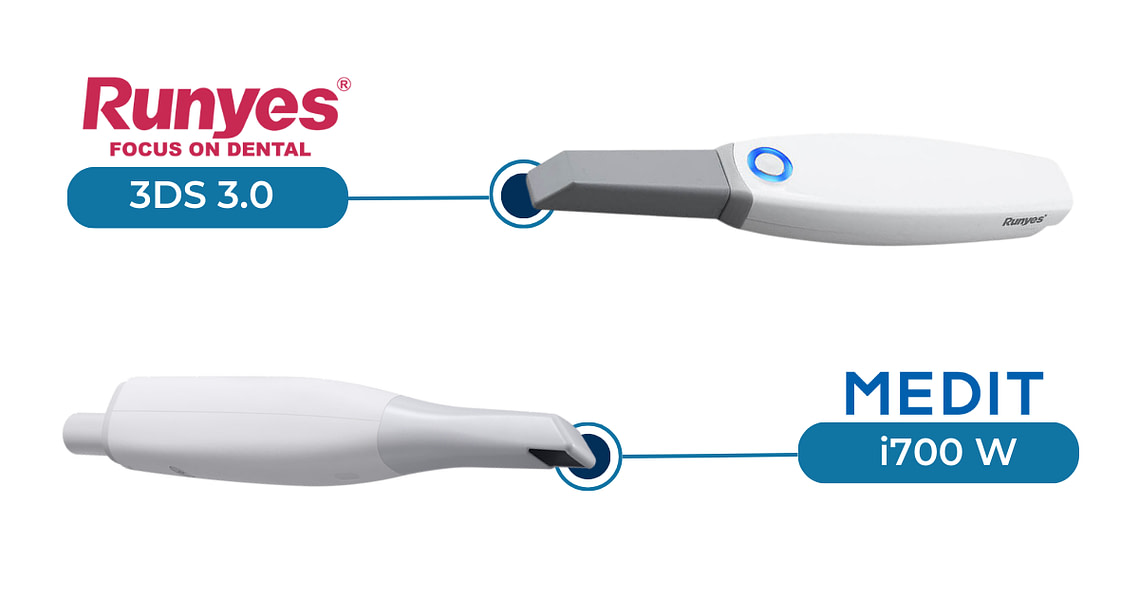
Scans in their Native Software
As we all know by now (or maybe you don’t as you’re new to intraoral scanning), every scanner captures color slightly differently depending on how accurately the scanner is able to pick up the light bouncing back off the prep and adjacent teeth.
Every intraoral scanner available comes with its native scanning software. Using AI, they often filter out scan irregularities such as movable soft tissues, cheeks, and tongue.
In the series of pictures below, you will see multiple images with a focus on the scan body and soft tissue, both in color and monochrome as previewed in their native software.
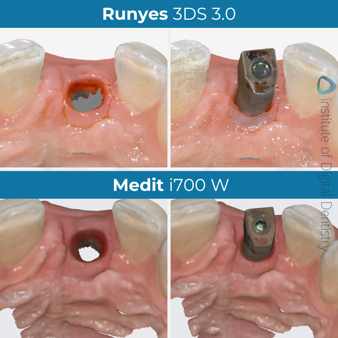
All scans were captured and exported in their highest, uncompressed definition, including the Medit i700 W, which was setup to scan in HD mode.
As you can see, there aren’t significant deviations in color, with only a slight difference in the scan brightness observed with the Runyes 3DS 3.0 scan.
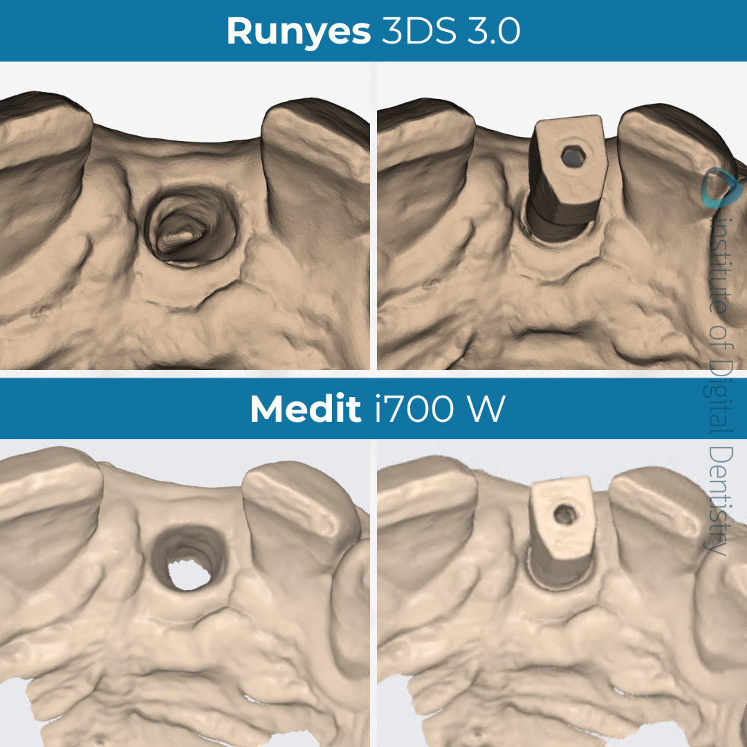
Monochrome scans can often offer a clearer perspective on the preparation quality and can be advised for identifying potential scan problems less visible in color.
Exported Scans in Third-Party Software
Most intraoral scanners consist of an open architecture to allow for scans to be exported and sent to labs. These scans are usually exported in three various formats - STL, PLY, or OBJ files.
Both IOS devices, Medit i700 W and Runyes 3DS 3.0 are capable of exporting scans in all three file formats - STL, PLY, and OBJ.
STL File Sizes Compared
Medit i700 W Scan Body
Medit i700 W Scan Tissue
Runyes 3DS 3.0 Scan Body
Runyes 3DS 3.0 Scan Tissue
PLY File Sizes Compared
Medit i700 W Scan Body
Medit i700 W Scan Tissue
Runyes 3DS 3.0 Scan Body
Runyes 3DS 3.0 Scan Tissue
OBJ File Sizes Compared
Medit i700 W Scan Body
Medit i700 W Scan Tissue
Runyes 3DS 3.0 Scan Body
Runyes 3DS 3.0 Scan Tissue
STL files export as monochromatic scans whereas OBJ and PLY files store color and texture. Not all intraoral scanners are able to export OBJ and/or PLY files whereas STL files are widely used across the market as a default setting. As we can see from the table above, OBJ file format had the largest MB size for Runyes 3DS 3.0 in both scanning surfaces (scan body and scan tissue). OBJ files can contain detailed information like color and texture as well as more complex geometric information such as curves and surfaces, like a scan body in this case. Because of this, it is preferable to use an OBJ file with more complex dental structures to store this type of information.
Labs use third-party CAD software (in our comparison’s case we used Medit Design) to preview the received STL, PLY, or OBJ files and create restorations based on these scans. By exporting the scans outside their native software, we are able to view the scans objectively without the customized color and optimized surface rendering of the individual scanner’s built-in software.
Below, you can also see the exported STL files in third-party software, Medit Design, with and without the tessellated mesh.
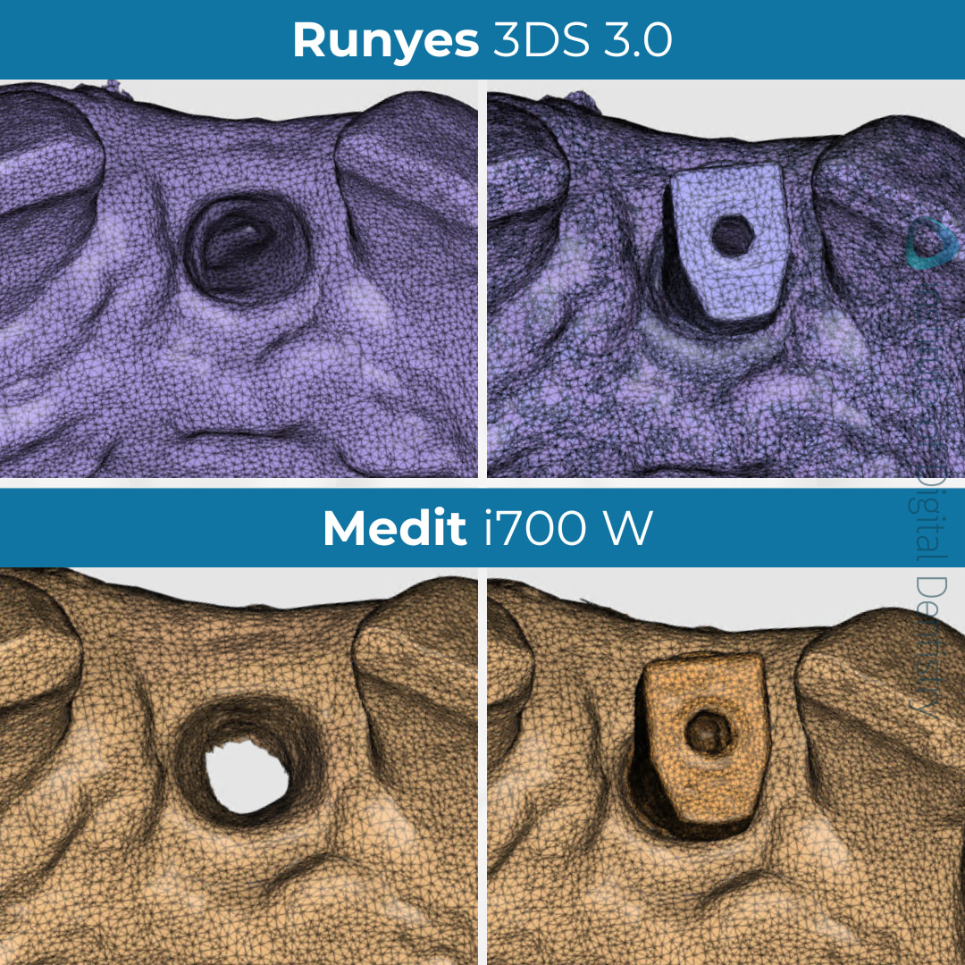
Tessellated mesh of Medit i700 W and Runyes 3DS 3.0 intraoral scans as viewed in Medit Design.
The comparison between the tessellated mesh of each scan is very slight but the Runyes 3DS scan seems to be more dense than the scan of the Medit i700 W. This could reflect on the scan size of the file formats however does not necessarily mean it is more accurate.
An additional tip for those using Medit intraoral scanners - when exporting your implant scans, you may notice that the Medit scan body scan appears to be more patchy and complete. This is a workflow issue, as there's a specific way to export such files from the Medit Link software. This is done by checking the "Combine Individual Mesh" option in the Export window before exporting the files.
Scan Accuracy
With Runyes as our point of reference, we can view the deviations of its scan to the Medit i700 W scan when aligned using Medit Design’s Deviation Display mode. Based on the colored deviation key, we can see that the scan meshes are -0.050 to +0.050 mm deviation compared to the scan taken with Runyes 3DS 3.0.
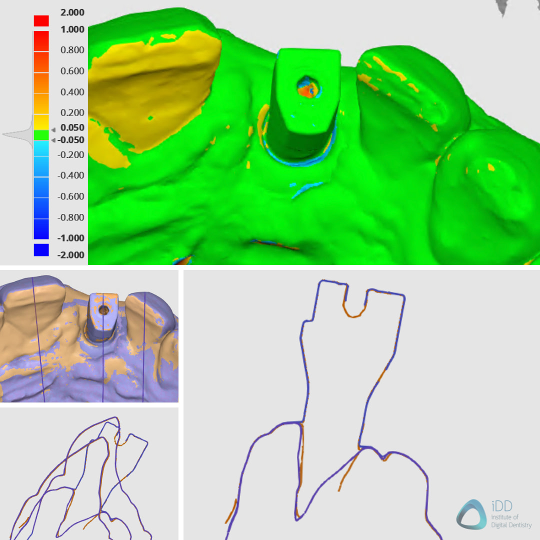
Deviation map of aligned scans, with Medit i700 W being our point of reference.
Within the same Display mode, we can view the aligned scans in a sectional view in which we can see minimal differences between the scans.
Conclusion
iDD objectively tests and reviews intraoral scanners to help other dental professionals form their opinions. We hope posts like this help answer some of your questions and help you make a more informed purchase.
That said, if there is anything else you would like us to post, let us know in the comments below.
Which IOS or type of cases would you like to see next?

Hi, I feel that the conclusions were missing, do you feel there is no difference? (without thinking about the software)
Yes, there was little to no clinical difference for a simple implant case like this.
That is the point. They all work. Any scanner can do this, especially when there is no blood or moving tissue. They all do the job.
People seem to think that the more they pay, the better accuracy they will get. This is not necessarily true.