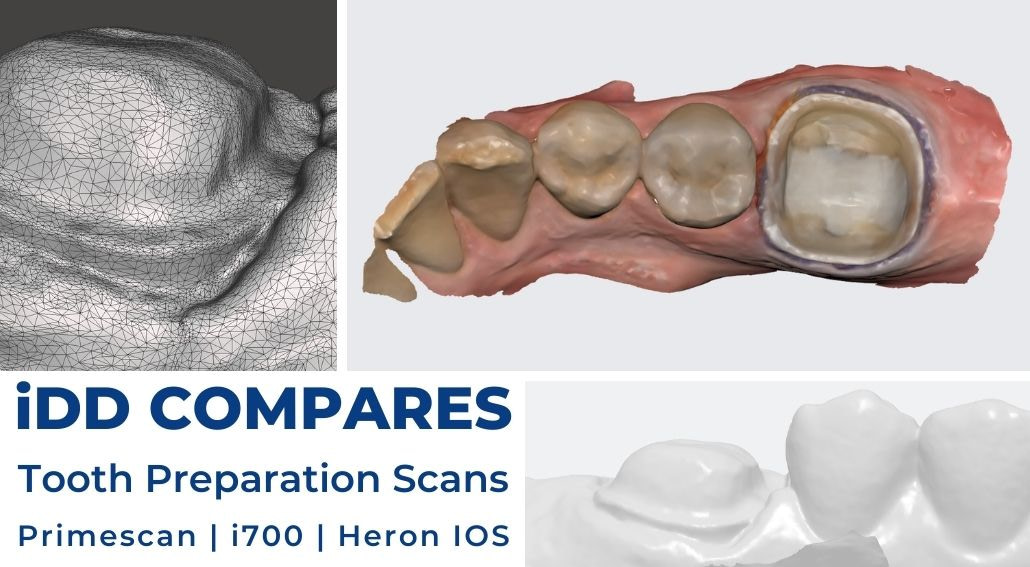
In this article, we are going to look at how relatively affordable intraoral scanners compare with one of the most expensive one on the market. I scanned the same patient, same preparation with three different scanners on the same day and these are the results.
Here at the Institute of Digital Dentistry, we often get asked whether the high-end intraoral scanners are really worth the investment or whether the “cheap” ones do the job and our answer is generally the same: it depends on what you want a scanner for.
Choosing the right intraoral scanner depends on multiple factors. Beside the initial investment cost, the most important thing to consider is whether you plan to bring the production in-house. If so, are you thinking of same-day workflows or involving a dental technician in some cases? Or maybe you are ok with leaving all the production to an external lab.
In this article, we have used scanners that could be considered by three types of dentists:
CEREC Primescan in combination with Primemill is our clinic’s favourite chairside system for same-day workflows. Fast, simple, and efficient but arguably too expensive for many.
Medit i700, a much cheaper alternative for dentists that like to effectively communicate with their lab but also want to get into other avenues of digital dentistry such as 3D printing – their free software and apps enable you to marginate cases, create printable models, showcase orthodontic and smile design simulations etc.
3DISC Heron IOS is a scanner that comes with no apps; therefore it might be considered by dentists that just want to scan and do not wish to delve into dental technology too much. It comes at a similar price to the Medit i700. It is simple to use but the software offers little more than exporting scans.
There are studies analysing the accuracy and trueness of intraoral scanners published every year. The Institute of Digital Dentistry is not a research facility, so we leave the scientific research to others. However, we are very fortunate to be in a position to test all intraoral scanners in a clinical environment, on real cases and real patients.
We have already voiced our opinions and shared our experiences in the reviews of each of these scanners. In this article, we want to show you how these scanners performed when scanning the same preparation on the same day. If you haven't read them already, click here for the CEREC Primescan review, Medit i700 review and 3DISC Heron IOS review.
Individual Scans in their Native Software
First, let’s look at what the scans look like in their native software, both in color (texture) preview and monochrome preview (below).


Every scanner opts for different processing of color and texture, which is what you see here. While the color view is helpful when assessing where the prep ends and gingiva starts, the monochrome makes it easier to spot scan issues such as holes, stitching, or even interferences caused by insufficient moisture control.
The quality of these previews is important for analyzing the scans before sending them. It becomes even more crucial when you start marginating cases within the same system (third-party CAD software generates colors differently).

Close-ups of the mesial-buccal margin as previewed in the native software of each intraoral scanner after processing.
As you can see in the picture above, the difference can be quite striking. Marginating preps might prove to be more difficult in one software than another, if they even provide the tools to do so.
Comparing Scan Export STL’s in a Third-Party Software
The real difference between the scans is then more obvious after you import the scans into third-party software. In this case, we used Meshmixer, a free CAD program by Autodesk.

We exported the scans in an STL format. The i700 files were exported with default settings, and the patient was not scanned with the HD mode on.
The Primescan and Medit files were exported in the highest definition, while the Heron IOS did not give us a quality or file size option. All were quadrant scans - I scanned the same area. That being said, this is the size of the exported files:

Unedited and untrimmed quadrant scans were captured on the day. Exported STL’s, viewed in Meshmixer.
In the following picture, you can see what arguably matters the most: the margin. On the left side of the picture, you can see the close-up of the same area in monochrome. On the right side, you can observe the tessellated mesh, which gives you an idea of the data density and it also explains the size of these files.

You can also see these close-ups in individual screenshots below - click to see them in higher resolution.


Conclusion
There is a noticeable difference between these scans, both within their native systems or a third-party CAD software.
From this case alone, it’s not conclusive whether there is any clinical significance of the different ways software produces digital impressions. Some are evidently more clear than others but does this impact margin placement and restoration fit?
More in-depth research in a controlled environment is definitely necessary to draw a proper conclusion whether this would impact the accuracy of margin placement.
This article is meant as a tool to help you decide which intraoral scanner might be better suited for your dental practice and its needs.
Given that the internal surface of the restoration normally has a "spacer", the clinical significance of intricate detail on the surface is likely to be nil. Marginal fit is obviously important but the differences are too small to notice, and, in all honesty, likely to be irrelevant due to the size of the milling bur. Other applications are unlikely to need this detail.
More relevant to me is the ability to generate an accurate representation of edentulous spans to prevent distortion.
Hello Duncan,
thank you for your comment, you made many good points and I believe you’re right. And yes, the long-span edentulous scans are proving to be the hardest for most scanners. Any time we scan such a case, we always use photogrammetry to ensure accurate results, no matter which IOS we use. We also plan to compare an edentulous case soon.
A very interesting comparison, thank you.
Thanks, David. Glad you liked it!
Which one do you suggest among three in-terms of scanning and Ortho simulation
Hi Dr Kiran,
In regards to scanning, they all perform sufficiently well. CEREC is arguably the best but it is more than double the cost of the others and doesn’t really make sense to me unless you go the whole workflow for chairside restorations.
In regards to ortho simulation, that’s easy. CEREC and Heron do not have any orthodontic simulations built-in, only Medit software does currently.
Check out our reviews of each scanner for more info:
https://instituteofdigitaldentistry.com/ios-reviews/cerec-primescan-and-primemill-review/
https://instituteofdigitaldentistry.com/cad-cam/heron/3disc-heron-intraoral-scanner-review
https://instituteofdigitaldentistry.com/ios-reviews/medit-i700-review/